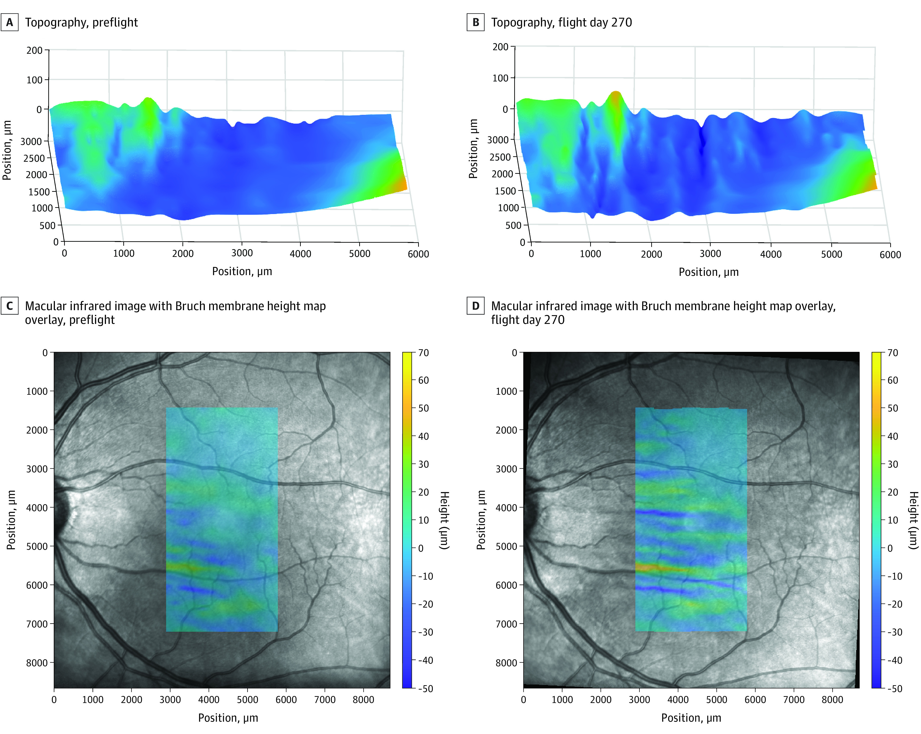
Figure 1. Optical Coherence Tomography Images of the Left Eye Show the Development of Macular Choroidal Folds in Flight.

Macular choroidal folds progressively worsened in participant 1 throughout the 1-year–long mission. Bruch membrane was segmented by a single expert grader and verified by a second expert. Fold metrics were quantified based on deviation from a nondeformed Bruch membrane layer using MATLAB software (MathWorks). A and B, Bruch membrane topography (left side corresponds with the inferior side of images in C and D). C and D, Macular infrared image with Bruch membrane heightmap overlay. See eFigure 1 in the Supplement for an example of Bruch membrane segmentation.
