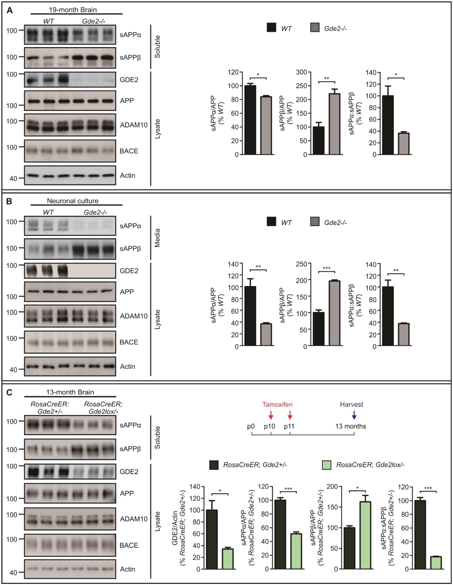
Fig. 3. GDE2 ablation reduces α-secretase and increases β-secretase APP cleavage.

A, Western blots and graphs quantifying sAPPα (sAPPα/APP *p=0.0122), sAPPβ (sAPPβ/APP **p=0.0071) and sAPPα:sAPPβ (*p=0.0201) in 19-month Gde2−/− cortices compared with WT. (n=3). B, Western blots and graphs quantifying sAPPα (sAPPα/APP **p=0.0097), sAPPβ (sAPPβ/APP ***p=0.0004) and sAPPα:sAPPβ (**p=0.0060) in DIV14 Gde2−/− cortical neurons compared with WT. (n=3). C, Western blots and graphs quantifying GDE2 expression (GDE2/Actin *p=0.0158), sAPPα (sAPPα/APP ***p=0.0004), sAPPβ (sAPPβ/APP *p=0.0176), and sAPPα:sAPPβ ratio (***p=6.69E-05) in 13-month RosaCreER;Gde2lox/− cortical lysates (n=3). Tamoxifen was injected on postnatal (p) days 10 and 11 in RosaCreER;Gde2+/− and RosaCreER;Gde2lox/− mice, and animals were harvested at 13-months of age. All graphs: Mean ± SEM, two-tailed unpaired Student’s t-test.
