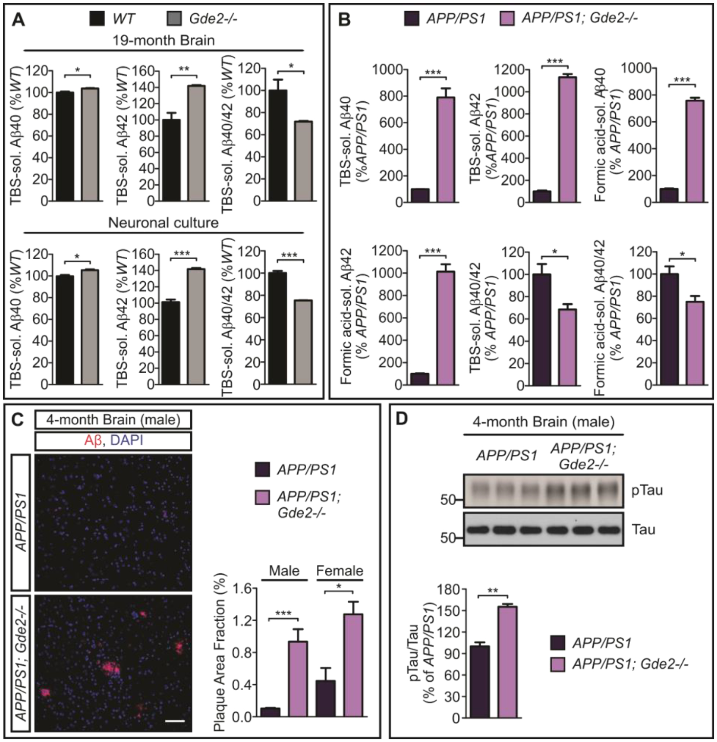
Fig. 4. GDE2 ablation promotes amyloidogenic APP processing.

A, ELISA quantifications of TBS-soluble Aβ40 (*p=0.0252), Aβ42 (**p=0.0088) and Aβ40/42 ratios (*p=0.0461) in Gde2−/− 19-month cortical extracts (n=3); and TBS-soluble Aβ40 (*p=0.0206), Aβ42 (***p=0.0003) and Aβ40/42 ratios (***p=0.0002) in Gde2−/− DIV14 cortical neuronal cultures (n=3). B, Quantification of ELISA in APP/PS1 and APP/PS1 Gde2−/− 4-month cortical extracts for soluble Aβ peptides (TBS-soluble Aβ40 ***p=0.0006; Aβ42 ***p=4.62E-06, n=3) and Aβ aggregates (Formic acid-soluble Aβ40 ***p=7.0E-06; Aβ42 ***p=0.0001, n=3), with associated Aβ40/42 ratios (TBS-soluble Aβ40/42 ratios *p=0.0399; Formic acid-soluble Aβ40/42 ratios *p=0.0444). C, Immunostaining of 4-month APP/PS1 and APP/PS1;Gde2−/− cortical sections with corresponding quantification of amyloid load [area fraction of 6E10-positive plaque (red)], male (***p=0.0007) and female (*p=0.0208). D, Western blots and quantification of phosphorylated tau (Ser202 and Thr205 pTau/Tau, **p=0.00127) in 4-month male APP/PS1 and APP/PS1;Gde2−/− cortical extracts (n=3). All graphs: Mean ± SEM, two-tailed unpaired Student’s t-test.
