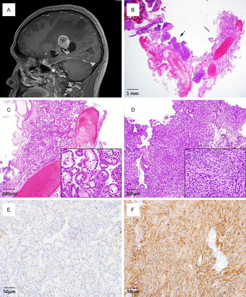
Figure 1.

(A) Brain MRI shows a well-defined and intense enhancing mass in the atrium of the right lateral ventricle. (B) The ventricular tumor comprises two distinct components: a choroid plexus papilloma (white arrow) and meningioma (black arrow). (C) In the choroid plexus papilloma, complex and delicate papillary fibrovascular fronds are covered by a single layer of uniform cuboidal-to-columnar epithelial cells. (D) Meningioma cells are arranged in lobules and are packed together in short fascicles, forming whorls and syncytial structures. (E) In the choroid plexus papilloma, tumor cells are negative for immunohistochemical staining of the epithelial membrane antigen (EMA, 1:100, clone GP1.4, Leica Biosystems: Novocastra, UK). (F) In contrast, tumor cells are diffusely and strongly positive for EMA in the meningioma component. (B-D, Hematoxylin and eosin; B, Scan view; C, D, × 40; inset, × 400; E, F, EMA, × 200).
