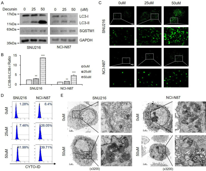
Figure 2.
Decursin induces autophagic alteration in GC cells. A, B. Decursin induced autophagosome formation in SNU216 and NCI-N87 cells, as revealed by conversion of LC3-I to LC3-II determined by western blotting. Full-length blots are presented in Supplementary Figure 6. C. Both cell lines were transfected with GFP-LC3 plasmid, and cells were treated with decursin for 24 h (×1000). Scale bar: 2 μm. D. Representative FACS data, showing accumulation of Cyto-ID after treatment with decursin. E. Effect of decursin on GC cell morphology. Representative TEM images are shown (×3200). Scale bar: 2 μm. The right panel shows the magnification of the boxed region. *P < 0.05; **P < 0.01; ***P < 0.001.

