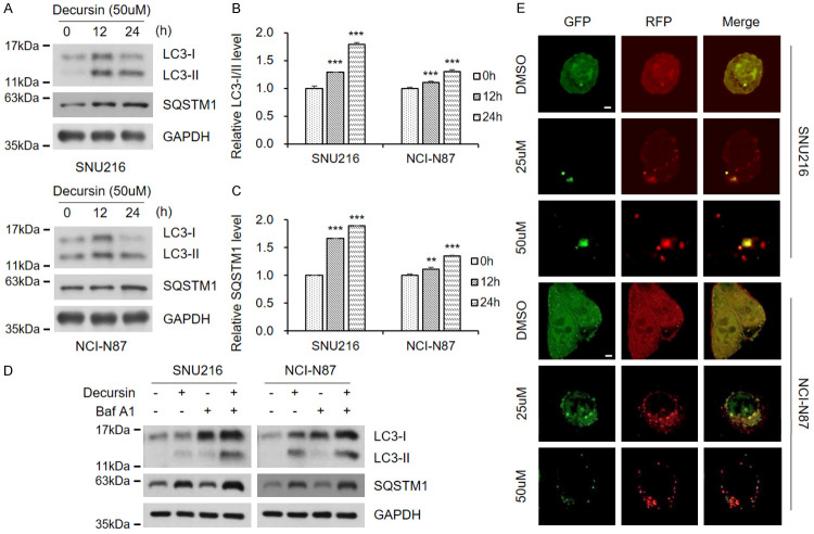
Figure 3.
Decursin interferes with autophagic flux in GC cells. A. Decursin concentration was fixed at 50 μM, and treatment time was adjusted. Full-length blots are presented in Supplementary Figure 6. B, C. Expression of LC3-II and SQSTM1. D. SNU216 and NCI-N87 cells were exposed to decursin (50 μM), bafilomycin (100 nM), or a combination of both drugs for 24 h. Full-length blots are presented in Supplementary Figure 6. E. Both GC cell lines were transfected with mRFP-GFP-LC3 and treated with decursin for another 24 h, then analyzed by fluorescence microscopy (×1000). Scale bar: 2 μm.

