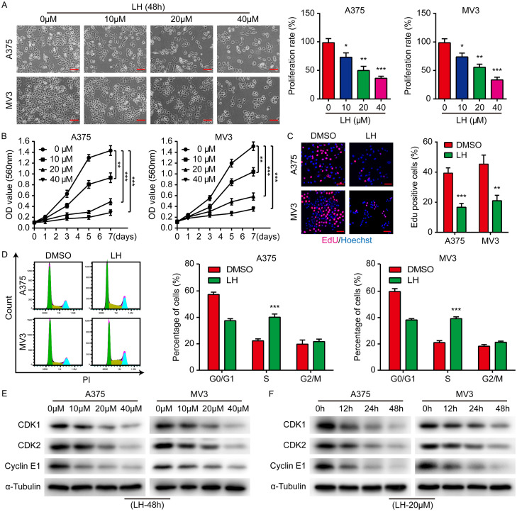
Figure 1.
Anti-proliferative effect of LH on melanoma cells. A. A375 and MV3 cells were treated with LH (0, 10, 20 and 40 μM) for 48 h and proliferation of the cells was observed. Cell percentage in 0 μM group is regarded as 100%. Scale bar, 100 μm. B. Under LH (0, 10, 20, and 40 μM) treatment, cell viability was measured by MTT assay on days 1, 3, 5 and 7. C. Percentage of Edu positive cells after LH (20 μM) treatment for 48 h. Scale bar, 50 μm. D. Cell cycle distribution of the cells after LH (20 μM) treatment for 48 h. E, F. The protein levels of CDK1, CDK2 and Cyclin E1 in LH treated cells were measured by western blotting. Mean ± SD; *P<0.05, **P<0.01, ***P<0.001.

