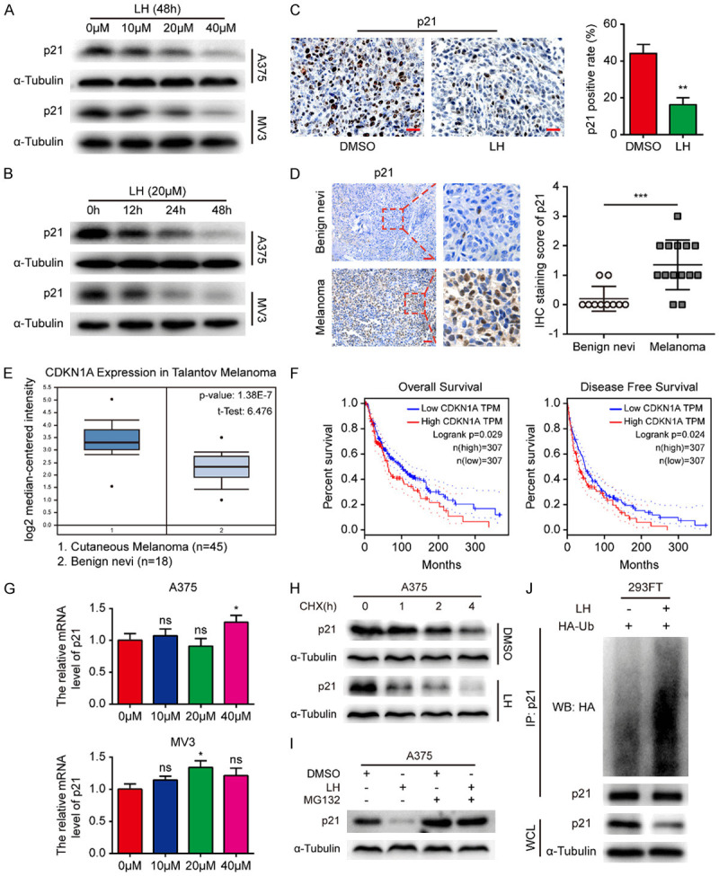
Figure 4.

High p21 expression indicates poor prognosis of melanoma and LH promotes ubiquitination degradation of p21. A, B. Expression of p21 in A375 and MV3 cells was detected by western blotting after LH treatment. C. Expression of p21 in the xenograft tumors from LH or DMSO treated mice model. IHC; Scale bar, 100 μm. D. Expression of p21 in human cutaneous melanoma (n = 14) was detected by IHC staining, benign nevi (n = 10) was as control. Scale bar, 200 μm. E. The CDKN1A level in cutaneous melanoma tissues (n = 45) compared with benign nevi tissues (n = 18) from Oncomine database. F. The association of CDKN1A gene with clinical outcomes (OS and DFS) of melanoma patients were analyzed through GEPIA database. G. The effect of LH on the expression of p21 mRNA in melanoma cells. H. The LH and DMSO pre-treated A375 cells were incubated with 10 μM CHX and collected at 0, 1, 2 and 4 h, cell lysates were immunoblotted with anti-p21 antibody. I. Cells were cultured in the absence or presence condition of 20 μM LH, the p21 protein level was detected after 20 μM MG132 treatment. J. The ubiquitination of p21 was detected in LH treated 293-FT cells. Mean ± SD; *P<0.05, **P<0.01, ***P<0.001; ns, not significant.
