
Figure 2.
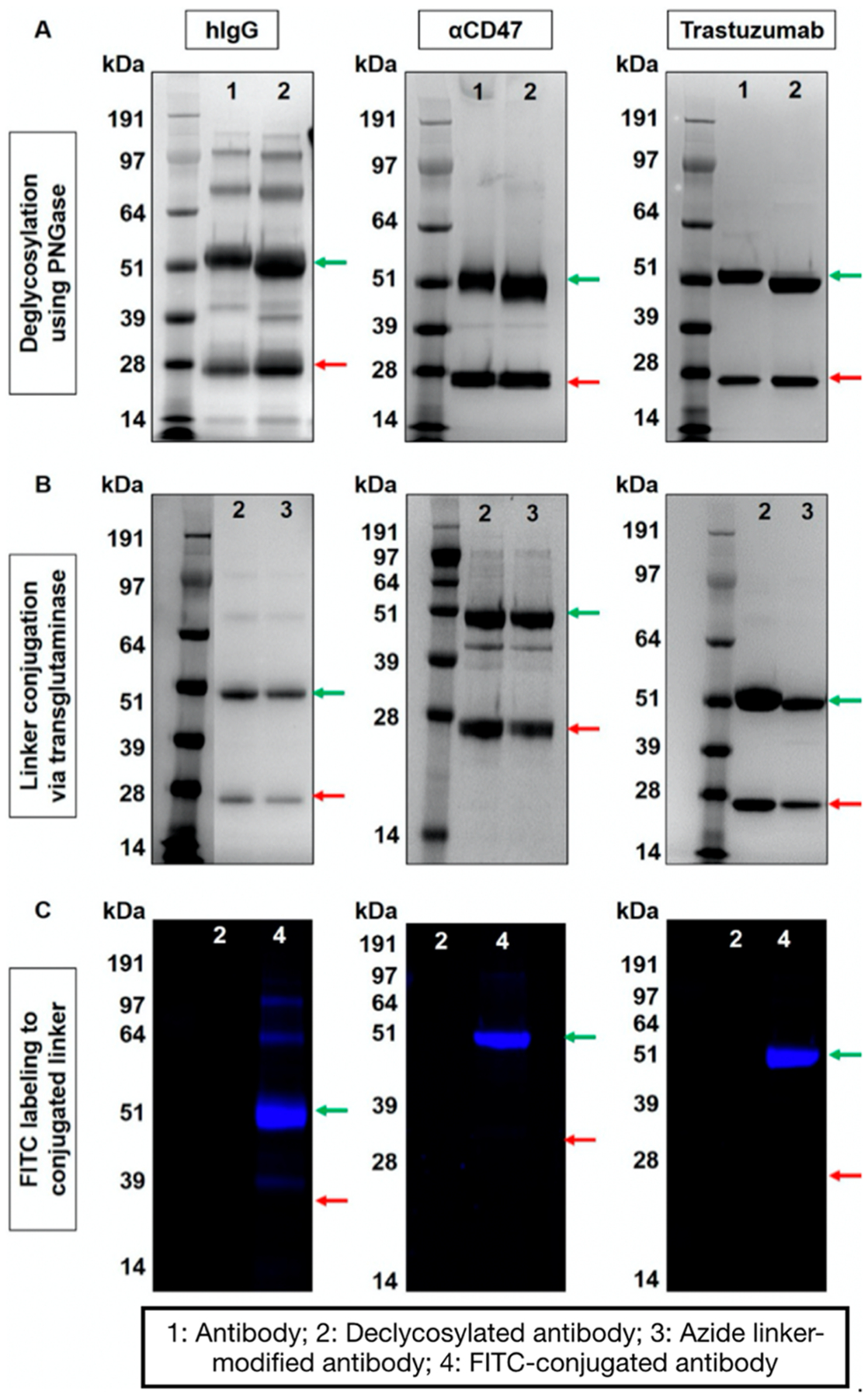
Characterization of site-specific antibody modification steps using gel electrophoresis. (A) IgGs pre- and postdeglycosylation using PNGase, (B) IgGs without and with azide-linker conjugated via transglutaminase, and (C) confirmation of the presence of the azide-linker via click reaction using FITC-PEG3-alkyne post transglutaminase reaction. Red and green arrows correspond to light and heavy chains of antibody (25 and 50 kDa), respectively.