
Figure 3.
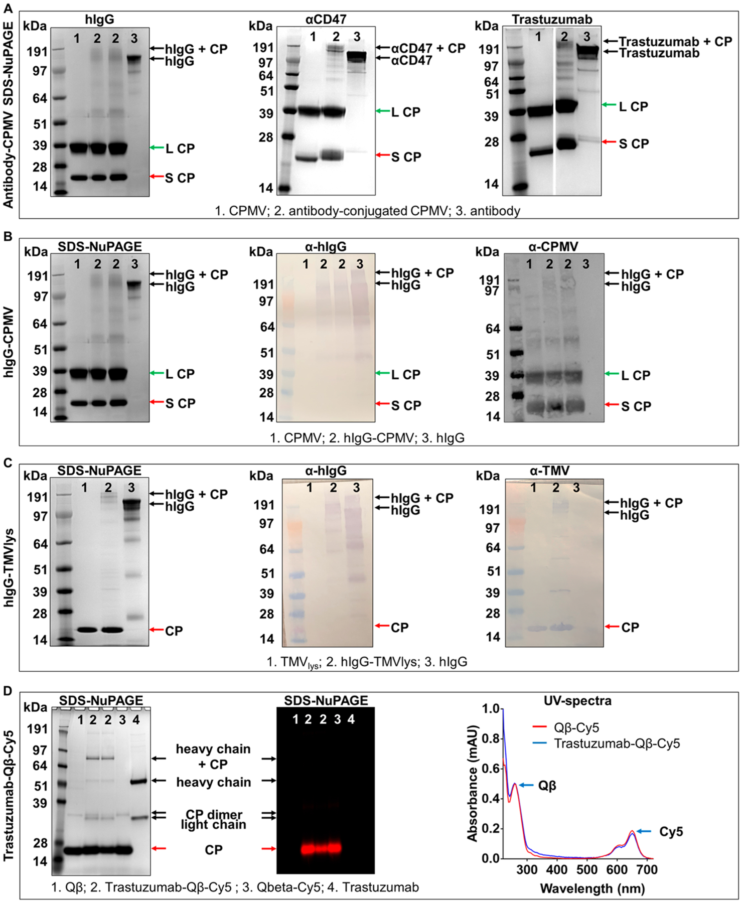
Conjugation of site-specifically modified antibodies to VNPs/VLPs via click chemistry. (A) SDS-NuPAGE gels of hIgG, αCD47, and Trastuzumab antibody conjugation to CPMV after Coomassie staining, (B) SDS-NuPAGE (left) of hIgG-CPMV and corresponding Western blot probed with anti-hIgG antibody (middle) and anti-CPMV antibody (right) (in Western blot additional high molecular weight bands appear in the CPMV sample; we attribute this to multimers of CPs; the Western blot is more sensitive than the SDS-NuPAGE and this is why these bands are not detected by SDS-NuPAGE). (C) SDS-NuPAGE (left) of hIgG-TMVlys and corresponding Western blot probed with anti-hIgG antibody (middle) and anti-TMVlys antibody (right). (D) SDS-NuPAGE of Cy5 labeled, Trastuzumab conjugated Qβ after Coomassie staining (left) and under red fluorescence (middle) to confirm Cy5 conjugation. Right panel corresponds to the UV-spectrum verification of Cy5 labeling onto Qβ.