
Figure 4.
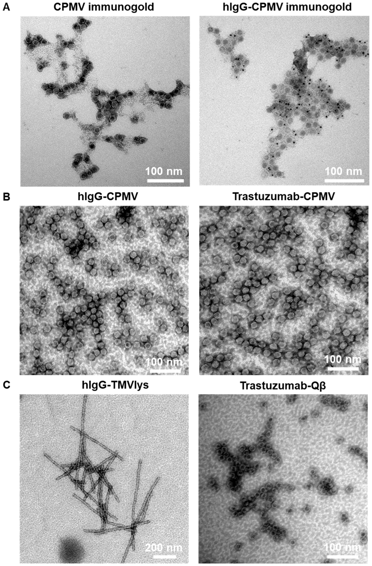
TEM images of antibody-conjugated VNPs. (A) Immunogold-staining of native CPMV and hIgG-CPMV hIgG. Six nm anti-hIgG labeled gold nanoparticles are visible as black dots only in the hIgG conjugated samples, (B) CPMV conjugated to hIgG (left) or Trastuzumab (right), (C) TMVlys conjugated to hIgG (left) and Qβ conjugated to Trastuzumab (right).