
Scheme 1. Fabrication of Antibody-Conjugated VNP1.
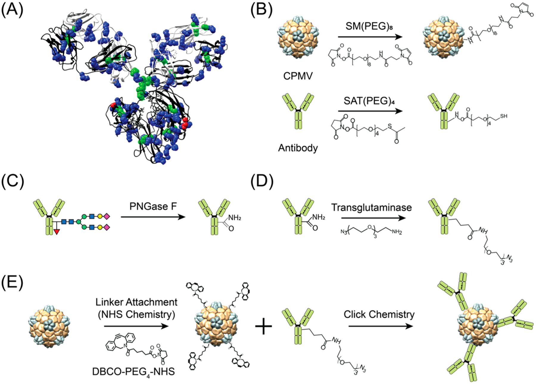
1(A) Structure of human IgG1 highlighting abundance of lysine (blue) and cysteine (green) and the location of Gln295 (red). (B) Lysine and cysteine targeted conjugation yields to heterogeneous antibody presentation. (C) Deglycosylation of Asn297 on heavy chains to make the adjacent Gln295 accessible, (D) site-specific attachment of a linker with azide (N3) group for conjugation via transglutaminase reaction, and (E) conjugation of azide-modified antibody to VNP/VLP. The structure of human IgG1 and cowpea mosaic virus (CPMV) were rendered using the UCSF Chimera software (PDB ID: 1IGY and 1NY7 for hIgG1 and CPMV, respectively).