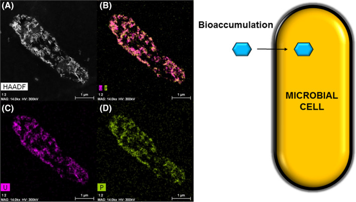
Fig. 4.

Example of intracellular accumulation of uranium (0.1 mM U(VI)) by Rhodosporidium toruloides as needle‐like structure localized at the inner cytoplasm membrane.
A–D. Micrograph and distribution analysis of uranium (purple) and phosphorus (green) from Gerber et al. (2018) and schematic representation of the bioaccumulation mechanism
