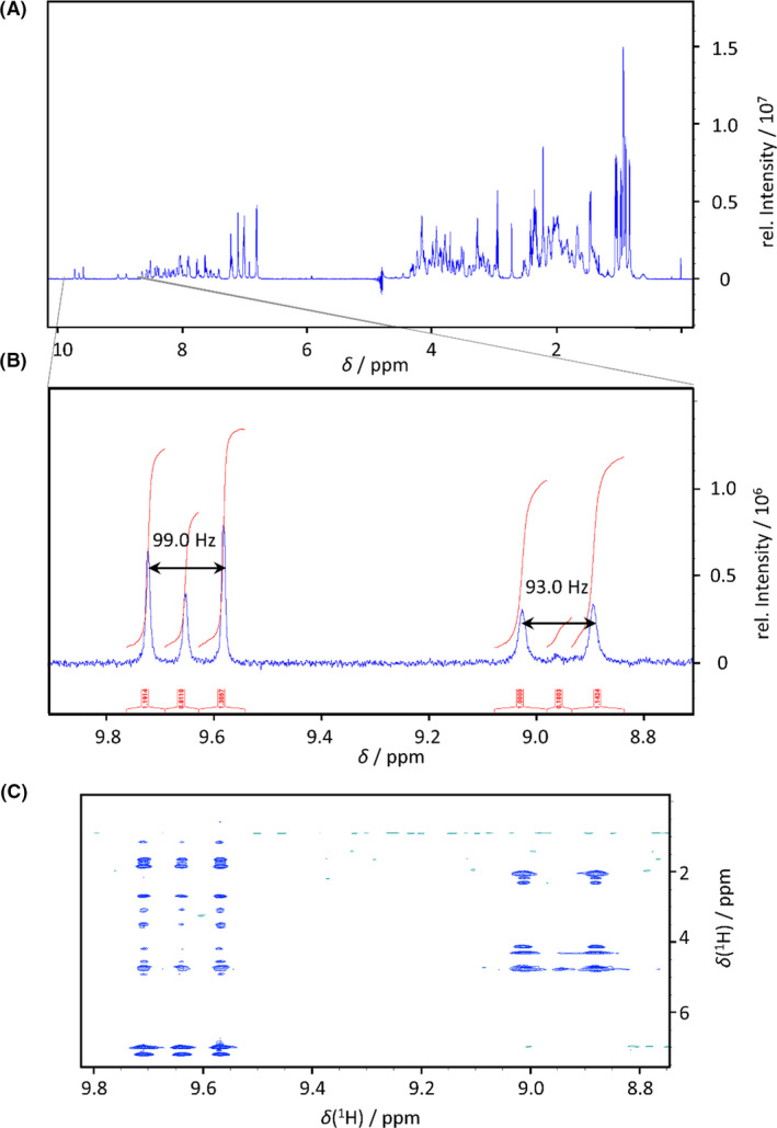
Fig. 5.

(A) 1H‐NMR spectrum of 15N‐E5 expressed in fermenter. (B) The zoom of the 1H‐NMR spectrum shows two different signal groups in the H(N) region: one is a typical resonance of backbone peptide H(N) (right) with a doublet with 1J‐coupling of 93.0 Hz (assigned to Glu3), while the other belongs to the Trp indole side chain H(N) with a 1J‐coupling of 99.0 Hz (left). (C) The zoom of the homonuclear 1H,1H NOESY spectrum proves that the three signals of Trp side chain and the two signals of the backbone belong to the same H(N) due to the same crosspeak pattern.
