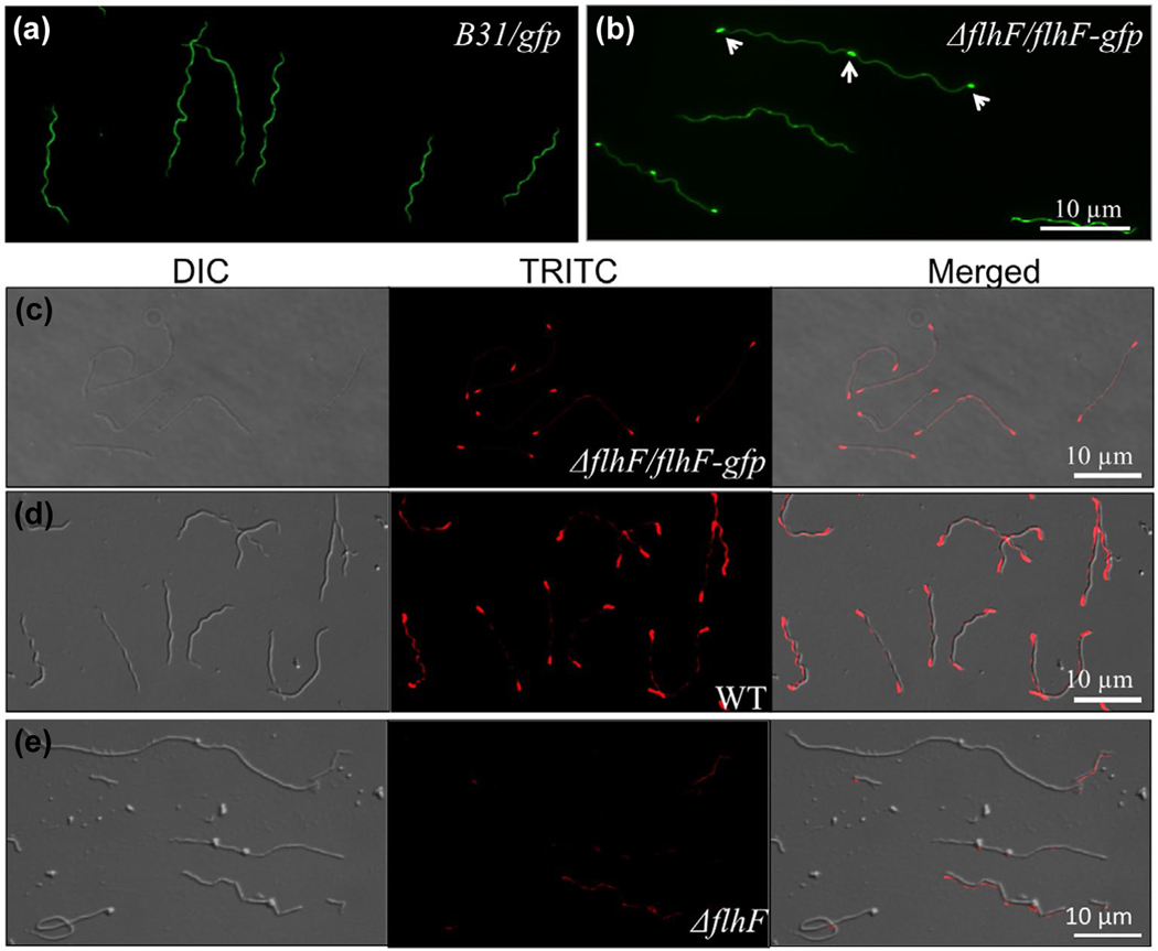
FIGURE 6.
Polar localization of FlhFBb in B. burgdorferi. (a) & (b) Fluorescence images of cells expressing GFP alone (a) or FlhFBb-GFP (b). The micrographs were taken under a fluorescence microscope (magnification, × 620) with a fluorescein isothiocyanate emission filter. (c-e) Immunofluorescence microscopic analysis of the ΔflhF/flhF-gfp (c), WT (d), and ΔflhF mutant (e) cells. Bacterial cells were fixed with methanol, stained with anti-FlhF and counterstained with anti-rat Texas Red-conjugated antibody, as previously described (Zhang, Liu, et al., 2012). The micrographs were taken under differential interference contrast (DIC) light and a fluorescence microscope with a tetramethylrhodamine isothiocyanate (TRITC) emission filter, and the resultant images were merged

