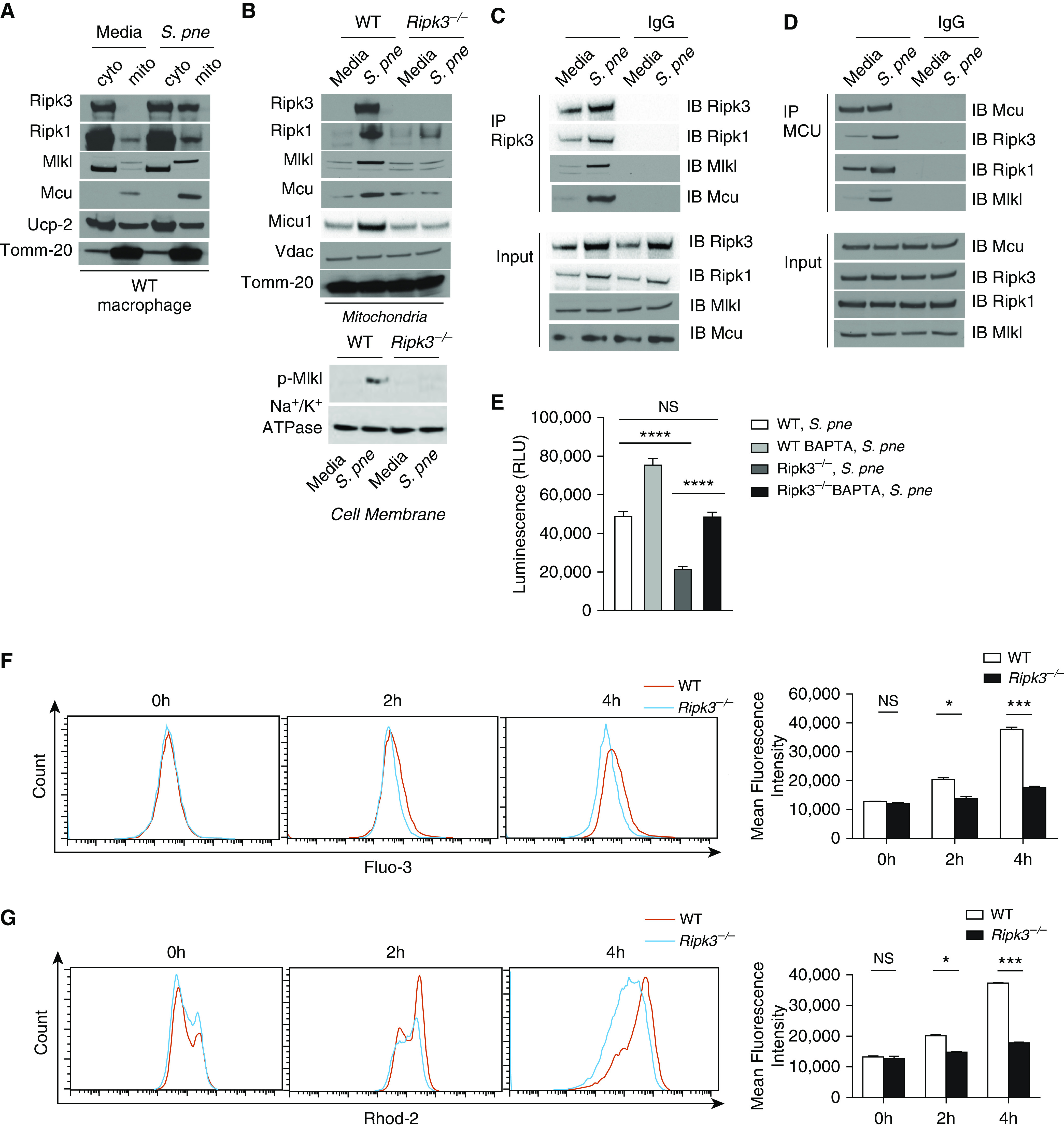
Figure 4.

RIPK3 interacts with MCU (mitochondrial calcium uniporter) in mitochondria. Macrophages derived from WT or Ripk3−/− mice were coincubated with S. pne, strain ATCC 6303 (MOI = 10), for 4 hours. (A) Mitochondrial (mito) and cyto fractions from WT macrophages were subjected to Western blot analysis for RIPK3, RIPK1, MLKL, MCU, and UCP2 (uncoupling protein 2). (B) Mitochondria lysates from WT or Ripk3−/− macrophages were subjected to Western blot analysis for RIPK3, RIPK1, MLKL, MCU, MICU1, and VDAC (voltage-dependent anion channel). Tomm-20 was the standard. (C) Coimmunoprecipitation of RIPK3 in total protein from naive or S. pne–infected cell lysates. (D) Coimmunoprecipitation of MCU in total protein from naive or S. pne–infected cell lysates. (E) WT and Ripk3−/− macrophages were pretreated with BAPTA-AM (10 μM, 20 min) before infection with S. pne (MOI = 10) for 4 hours. Cell viability was assessed using the Real Time Glo Viability assay, with decreased luminescence being indicative of cell death. ****P < 0.0001. (F–G) Macrophages derived from WT or Ripk3−/− mice were coincubated with S. pne (MOI = 10) for indicated hours. (F) Cells were loaded with 5 μM Fluo-3, and the intracellular calcium fluorescence was detected by flow cytometric analysis. (G) Cells were loaded with 5 μM Rhod-2, and the mito calcium fluorescence was detected by flow cytometric analysis. *P < 0.05 and ***P < 0.001 by two-way ANOVA test. Data are presented as mean ± SEM. Technical and biological replicates for experiments, N = 3–5. Experiments were repeated at least three times, with representative findings shown. BAPTA = 1,2-bis(2-aminophenoxy)ethane N,N,N′,N′-tetraacetic acid; cyto = cytosolic; RLU = relative light units.
