
Figure 7.
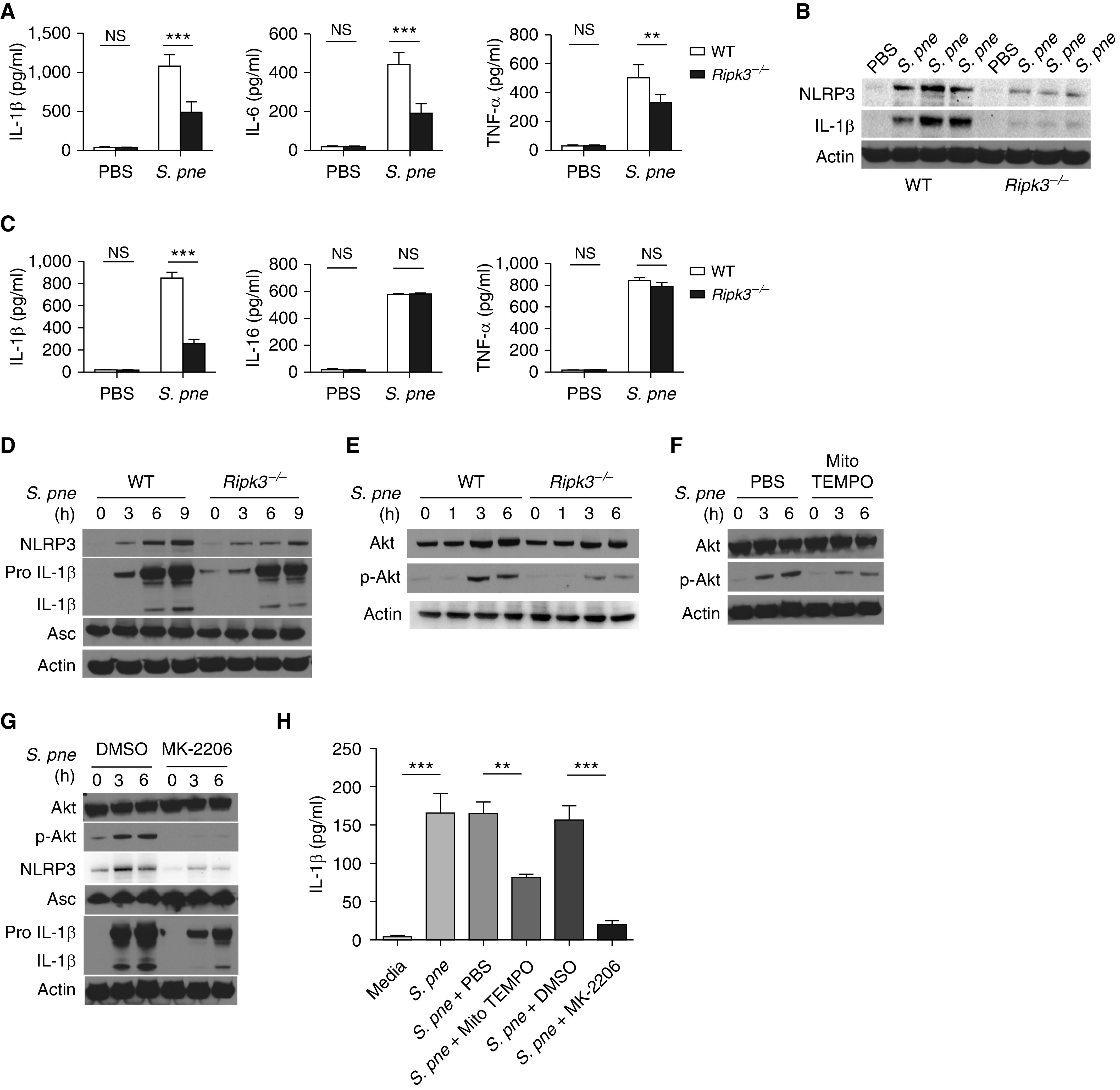
RIPK3 initiates NLRP3 inflammasome activation in response to S. pne infection. (A–B) WT or Ripk3−/− mice were intranasally instilled with 107 S. pne (ATCC 6303) and killed at 1 day after infection. (A) Cytokine concentrations were detected in the supernatants of lung tissue homogenates. (B) Pulmonary expression of NLRP3, IL-1β, and β-actin were examined by Western blot analysis in total protein from lung homogenates. (C–D) WT or Ripk3−/− macrophages were coincubated with S. pne (MOI = 10) for 18 hours. (C) Cytokine concentrations were detected in the culture supernatant. (D) NLRP3, Pro IL-1β, IL-1β, ASC, and β-actin expression were determined by Western blot analysis in total protein of cell lysis. (E–H) WT or Ripk3−/− macrophages were coincubated with S. pne (MOI = 10) for the indicated time. (E) Akt, phospho-Akt, and β-actin expression were determined by Western blot analysis in total protein of cell lysates. (F) WT macrophages were coincubated with S. pne (MOI = 10) with/without treatment of 20 μM MitoTEMPO for the indicated time. Akt, phospho-Akt, and β-actin expression were determined by Western blot analysis in total protein of cell lysates. (G) WT macrophages were coincubated with S. pne (MOI = 10) with/without treatment of 5 μM MK-2206 for the indicated time. Akt, phospho-Akt, NLRP3, Pro IL-1β, IL-1β, Asc, and β-actin expression were determined by Western blot analysis in total protein of cell lysates. (H) IL-1β concentrations were measure by ELISA in the culture supernatant of WT macrophages coincubated with S. pne (MOI = 10) for 18 hours with/without 20 μM MitoTEMPO or 5 μM MK-2206 treatment. **P < 0.01 and ***P < 0.001 by one-way ANOVA test. Data are presented as mean ± SEM. Densitometric analysis of all Western blots is included in Figure E5. Technical and biological replicates for experiments, N = 3–5. Experiments were repeated at least three times, with representative findings shown.