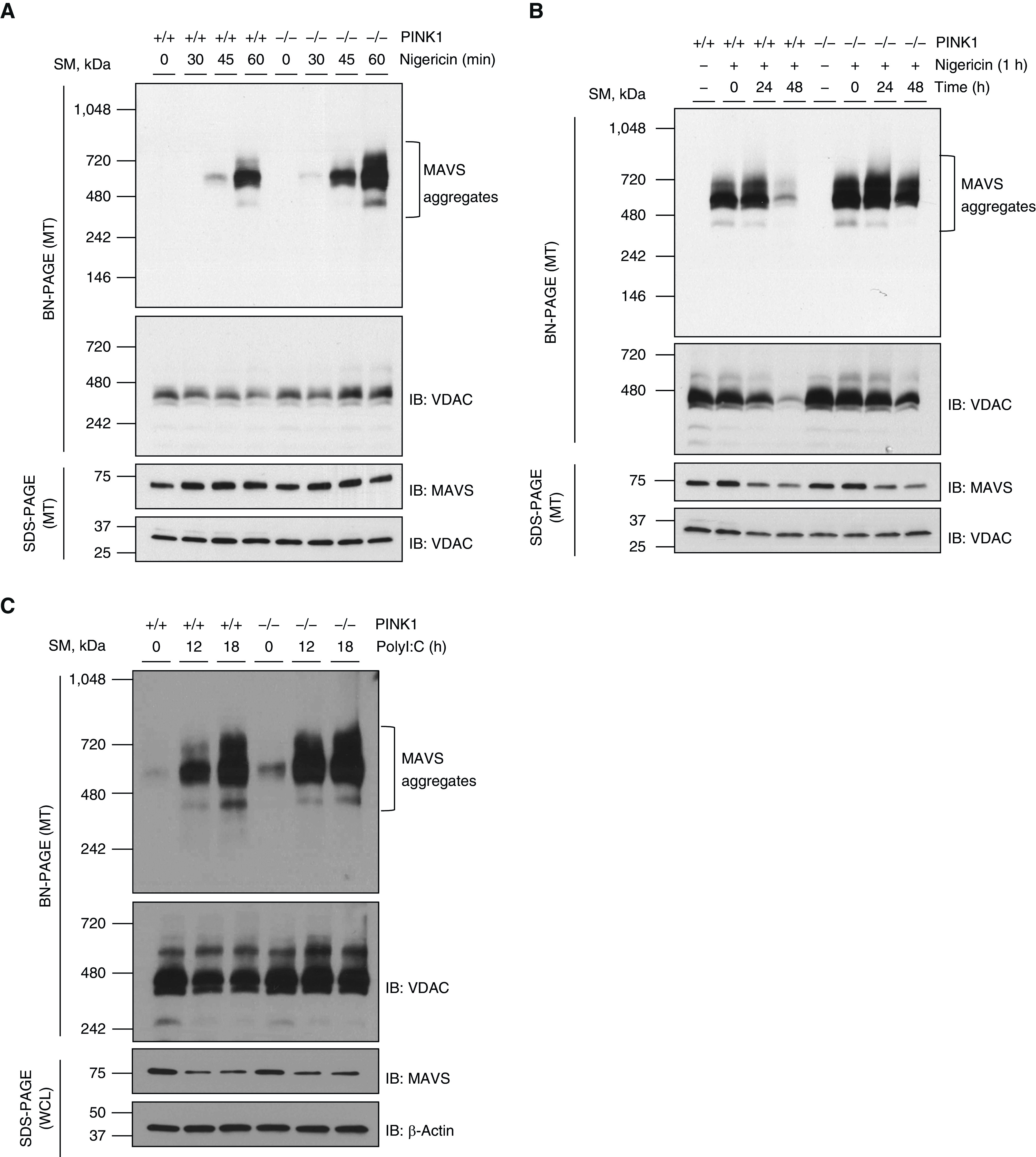
Figure 2.

The augmentation of MAVS aggregation in the absence of PINK1. (A) Mouse embryonic fibroblasts (MEFs) from wild-type (WT) and PINK1−/− mice were stimulated with 5 μM of nigericin for indicated time. MTs were separated and loaded to BN-PAGE. The MAVS aggregation was analyzed by Western blot. (B) MEFs from WT and PINK1−/− mice were stimulated with 5 μM of nigericin for 1 hour. Then, cells were washed with serum-free Dulbecco’s modified Eagle medium (DMEM) and maintained with 10% FBS/DMEM for indicated time. MTs were separated and loaded to BN-PAGE. The MAVS aggregation was analyzed by Western blot. (C) MEFs from WT and PINK1−/− mice were stimulated with 10 μg/ml of high molecular weight polyI:C for the indicated time. MTs and WCLs were separated and loaded to BN-PAGE and SDS-PAGE, respectively. The MAVS aggregation was analyzed by Western blot. VDAC and β-actin were used as loading controls of MTs and WCLs, respectively. All experiments are repeated at least three times. Representative results are shown. PolyI:C = polyinosinic–polycytidylic acid.
