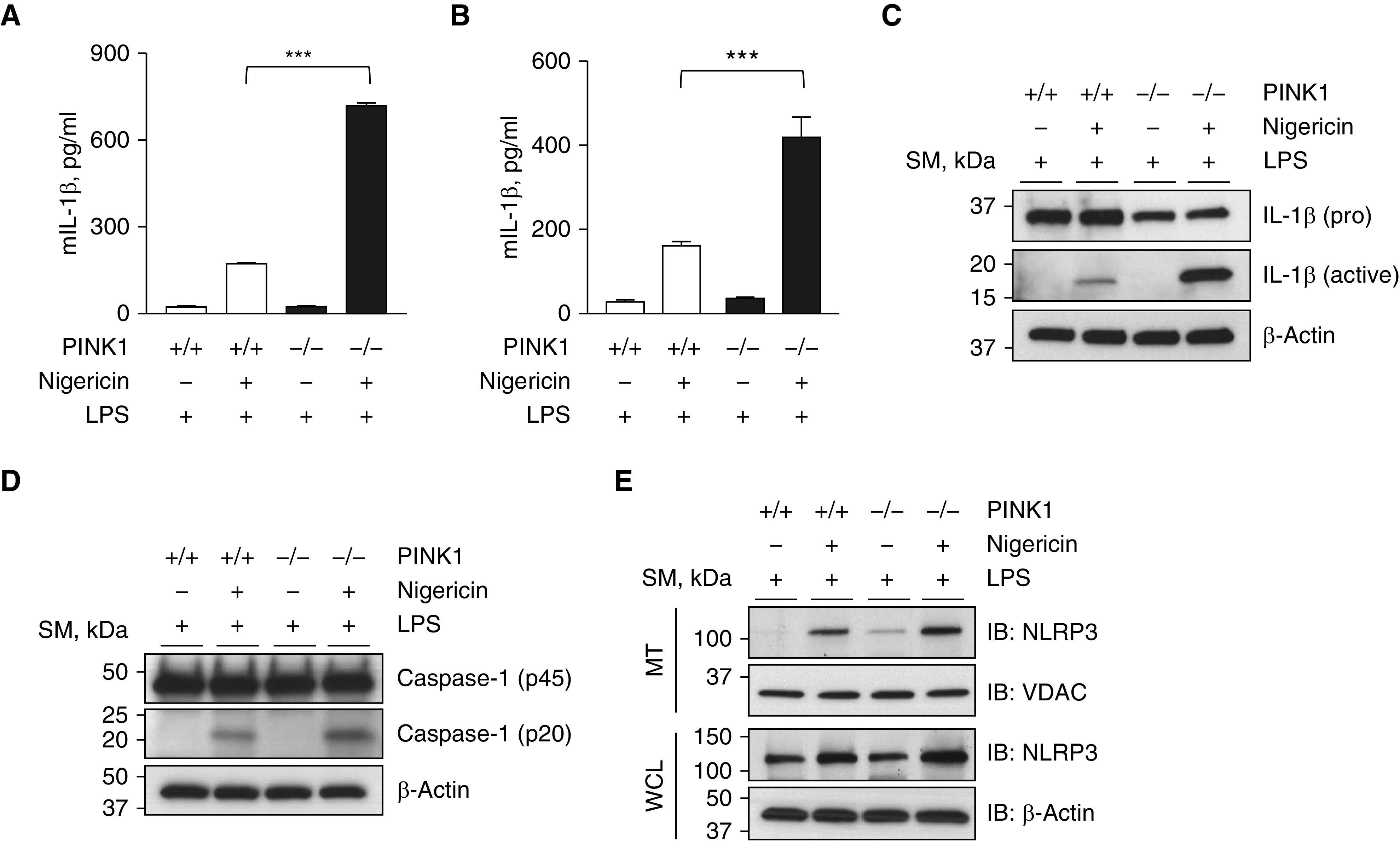
Figure 5.

Enhancement of the MAVS-mediated inflammasome signaling in the absence of PINK1. (A) MEFs from WT and PINK1−/− mice were primed with 1 μg/ml of LPS for 4 hours and then stimulated with 5 μM of nigericin for 1 hour. The supernatant was collected, and the amount of IL-1β was measured by ELISA. (B–E) BMDMs from WT and PINK1−/− mice were primed with 1 μg/ml of LPS for 4 hours and then stimulated with 5 μM of nigericin for 1 hour. (B) The supernatant was collected, and the IL-1β amount was measured by ELISA. (C and D) The amounts of pro forms and active forms of IL-1β (C) and caspase-1 (D), respectively, in cell lysates from BMDMs were evaluated by Western blot analysis. (E) Mitochondrial fractions from BMDMs were loaded to SDS-PAGE, and the recruitment of NLRP3 inflammasome on mitochondria was evaluated by Western blot analysis. VDAC and β-actin were used as loading controls of mitochondria and WCLs, respectively. All experiments are repeated at least three times, and representative results are shown. Means ± SD. ***P < 0.001. mIL-1β = murine IL-1β; NLRP3 = NOD-like receptor family pyrin domain-containing 3.
