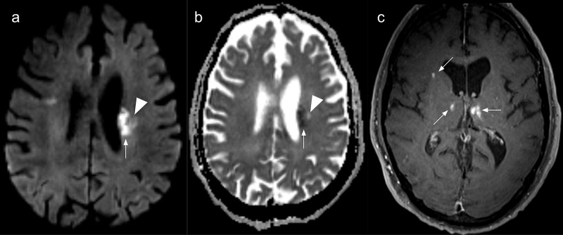
Figure 1. Brain magnetic resonance imaging.
Multiple and bilateral ischemic lesions in various evolution stages. (a) Axial diffusion-weighted image and (b) apparent diffusion coefficient (ADC) map showing an acute periventricular ischemic lesion (arrows). A chronic lesion is also seen as a facilitated diffusion area (arrowheads); (c) Axial contrast-enhanced T1-weighted image depicting enhancing subacute ischemic in both thalami and the right corona radiata.

