
Fig. 5.
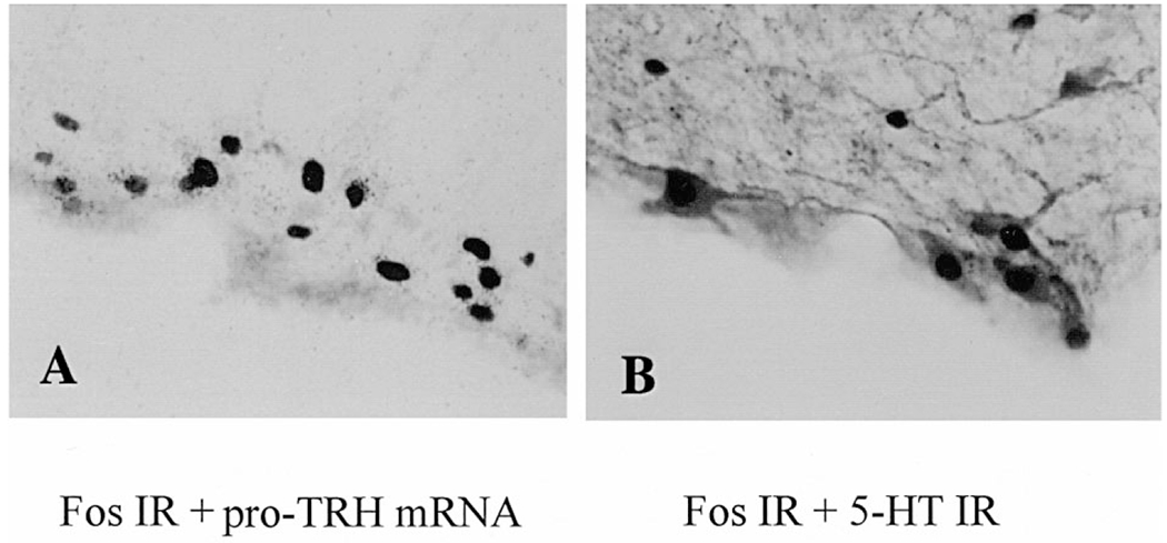
Double labeling of Fos-positive cells in TRH-and 5-HT-synthesizing neurons in the PPR in semi-restrained rats exposed to cold (4°C) for 2 h. (A) Combined Fos immunohistochemistry and pro-TRH mRNA in situ hybridization in bright field (interaural −3.30 mm, × 560). Fos-IR is shown as dark reaction product in the nuclei and pro-TRH mRNA appears as silver grains in the cytoplasm. (B) Double immunohistochemical staining for Fos-and 5-HT-IR (interaural −3.72, × 600). Fos-IR is shown as dark reaction product in the nuclei and 5-HT-IR as light/dark reaction product in the cytoplasm.