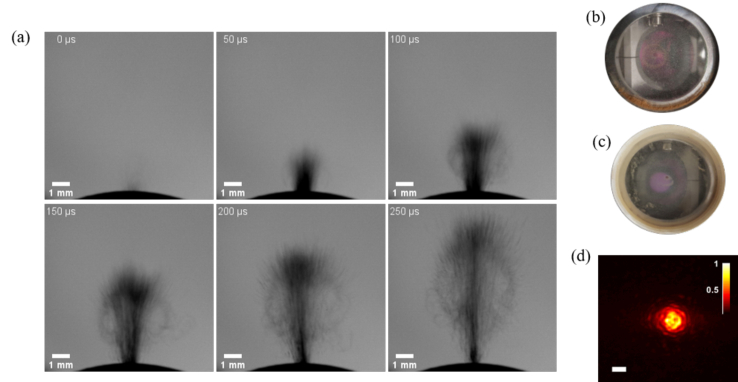
Fig. 7.
(a) Debris formation over time, from the onset of an Er:YAG laser pulse onto hard tissue. These images are captured using shadowgraphy method. (b)-(c) Debris accumulated on the sapphire. (d) Intensity profile of the Gaussian beam after passing through the windows covered with debris (scalebar = 200 µm).

