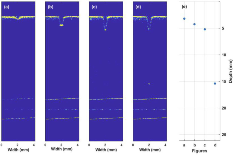
Fig. 11.
Selected images during a laser ablation process of bone collected using the long-range and extended DOF OCT system. (a)-(d) real-time depth monitoring of laser ablation of the femoral bone. (d) In this image, the ablative laser cut through the cortical line of the femur bone and reached the surface of the cortical bone in the medullary cavity. (e) The measured depth of cuts within the imaging range of the OCT system.

