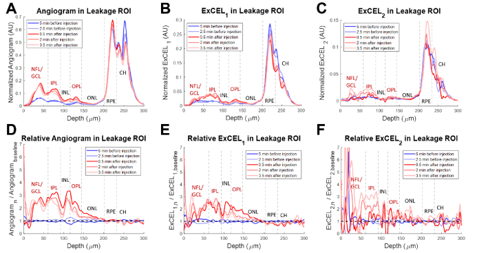
Fig. 5.
Depth profiles from a VLDLR mouse eye for the traditional angiogram (A), ExCEL1 (B), and ExCEL2 (C) signals averaged over the 100 µm × 100 µm leakage region shown in Fig. 3(B) (yellow box). Relative changes in signals (D-F) were also measured by dividing by the mean baseline profile. Blue profiles were acquired before Intralipid injection and red profiles were acquired after injection. NFL – Nerve Fiber Layer, GCL – Ganglion Cell Layer, IPL – Inner Plexiform Layer, INL – Inner Nuclear Layer, OPL – Outer Plexiform Layer, ONL – Outer Nuclear Layer, RPE – Retinal Pigment Epithelium, CH – choroid. Vascular layers of the inner retina are labeled in red.

