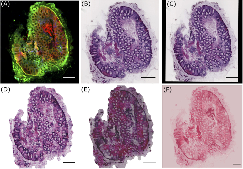
Fig. 1.
(A) shows a pre-processed NLM image with CARS, TPEF and SHG as the red, green and blue channel, respectively, (B) visualizes histopathologically stained H&E image (or unregistered H&E image) used for unsupervised pseudo-stain H&E model, (C) depicts a registered H&E image used for supervised pseudo-stain H&E model. The image in (C) shows the registration effect, which is filled with zeros. The images in (D), (E) and (F) are computationally stained H&E images with the supervised, unsupervised approach and method used in reference [12], respectively. All images are downscaled to 20% of the original size for clarity. The scale bar in all images represents 100 m.

