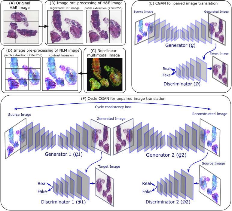
Fig. 2.
(A) is a histopathologically stained H&E image, (B) shows the image pre-processing of a histopathologically stained H&E image including image registration and patch extraction of size 256256, (C) is a corresponding NLM image, (D) visualizes the contrast inversion of the NLM image followed by patch extraction of size 256256, (E) shows a CGAN model for paired image translation which utilizes the registered histopathologically stained H&E images and contrast inverted NLM images, (F) depicts a cycle CGAN model for unpaired image translation using unregistered histopathologically stained H&E images and contrast inverted NLM images.

