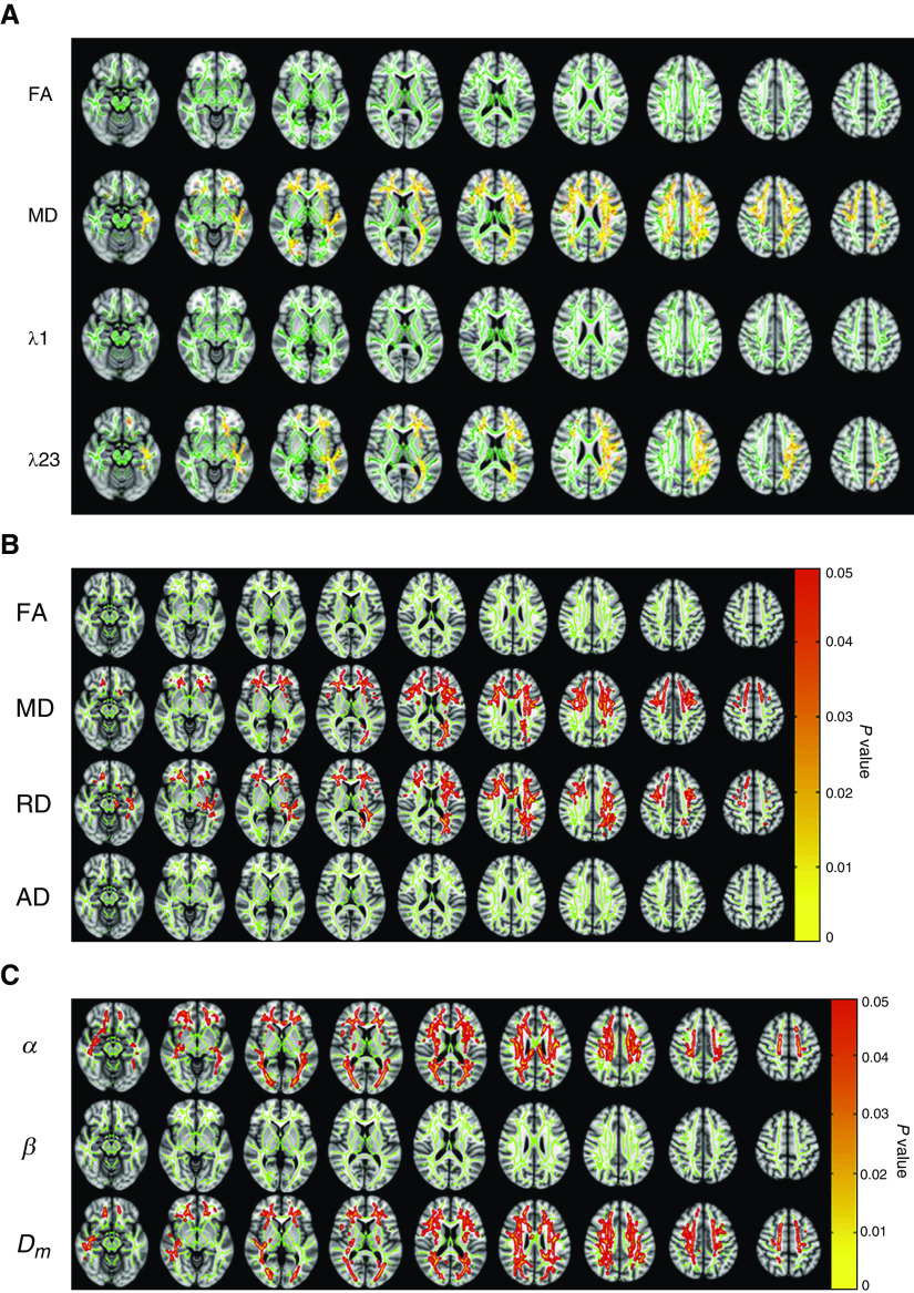
Figure 2.
White matter changes in patients with excessive daytime sleepiness associated with obstructive sleep apnea (OSA). (A) Diffusion tensor imaging (DTI) results showing possible white matter alterations in DTI metrics including FA, MD, λ1 (AD), and λ23 (RD) between sleepy and nonsleepy patients with OSA. Green: mean FA skeleton (threshold = 0.2) without significant change. Red-yellow: fibers with increased DTI metrics in the sleepy group when compared to the nonsleepy group (P < 0.05) (32). (B) Results from DTI whole-brain analysis based on FA, MD, λ23 (RD), and λ1 (AD) showing the presence or absence of differences between sleepy and nonsleepy patients with OSA. Age was included as a covariate in the group analyses for all parameters. Green: mean fractional anisotropy (FA) skeleton (threshold = 0.2) without significant change. Red-yellow: voxels with significantly increased parameters values in the sleepy group as compared to the nonsleepy group with P < 0.05 as shown in the color bar (33). (C) Whole-brain α, β, and Dm maps showing the presence or absence of differences between sleepy and nonsleepy patients with OSA (33). (A) Reprinted with permission of John Wiley & Sons. Xiong Y, et al. Brain white matter changes in CPAP-treated obstructive sleep apnea patients with residual sleepiness. J Magn Reson Imaging. 2017;45(5):1371–1378. Copyright © 2016 International Society for Magnetic Resonance in Medicine. (B and C) Reprinted from Zhang J, et al. White matter structural differences in OSA patients experiencing residual daytime sleepiness with high CPAP use: a non-Gaussian diffusion MRI study. Sleep Med. 2019;53:51–59. doi:10.1016/j.sleep.2018.09.011, with permission from Elsevier. AD = axial diffusivity; Dm = anomalous diffusion coefficient; FA = fractional anisotropy; MD = mean diffusivity; RD = radial diffusivity.

