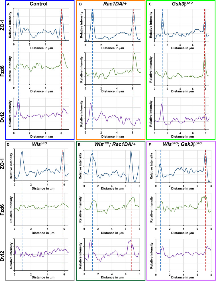
FIGURE 7.
Linescan analysis of junctional localization of Fzd6 and Dvl2 in outer hair cells. (A–F) Representative linescans of individual OHCs from E18.5 wild-type control (A), Rac1DA/+ (B), Gsk3βcKO (C), WlscKO (D), WlscKO; Rac1DA/+ (E), and WlscKO; Gsk3βcKO (F) OC stained for ZO-1, Fzd6, and Dvl2. For each genotype, a line was drawn parallel to the medial–lateral axis bisecting the OHC. Intensity profiles of each image channel were aligned along the distance axis. The lateral and medial junctions of the OHC were identified by peaks of ZO-1 staining and indicated by the blue and red dashed lines, respectively. Junctional Fzd6 and Dvl2 staining was defined by peaks in close proximity to the lateral or medial borders of the OHC.

