Figure 4.
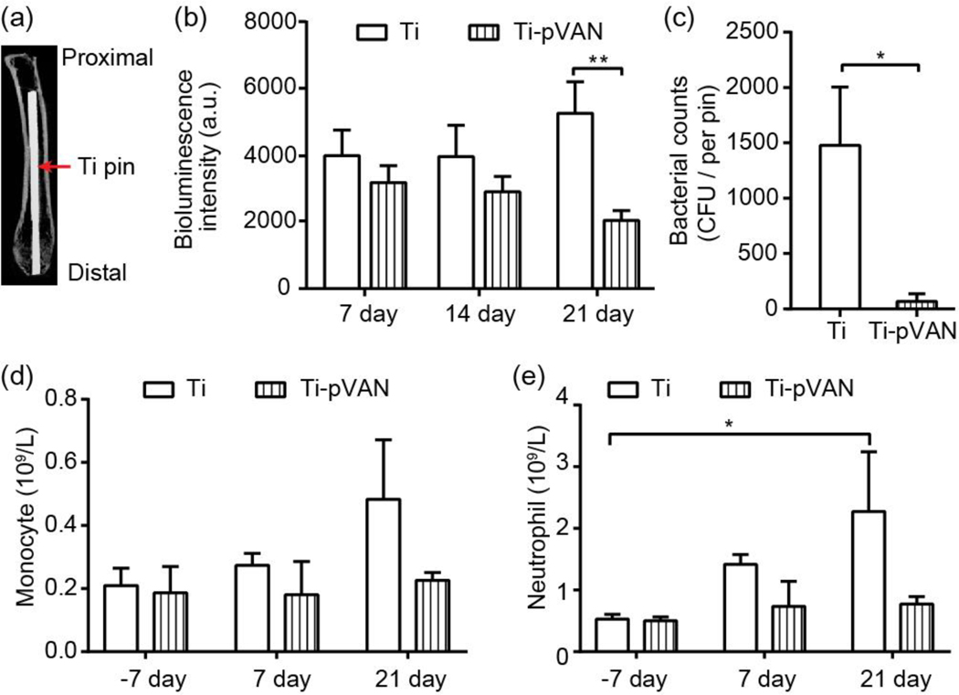
Ti-pVAN pins suppress S. aureus colonization in infected mouse femoral canal and mitigate infection in vivo. (a) μ-CT image confirming the proper insertion of an IM Ti6Al4V pin in mouse femoral canal. (b) Longitudinal bioluminescence monitoring of mouse femurs injected with 40 CFU S. aureus followed by Ti6Al4V or Ti-pVAN pin insertion (n=8). **P<0.01 (Two-way ANOVA). (c) S. aureus recovery from 21-day explanted pins (n=7), *p<0.05 (Student t test). Peripheral blood counts of monocytes (d) and neutrophils (e) before (−7 day) and after surgery (7 and 21 days; n=3). *p<0.05 (Two-way ANOVA).

