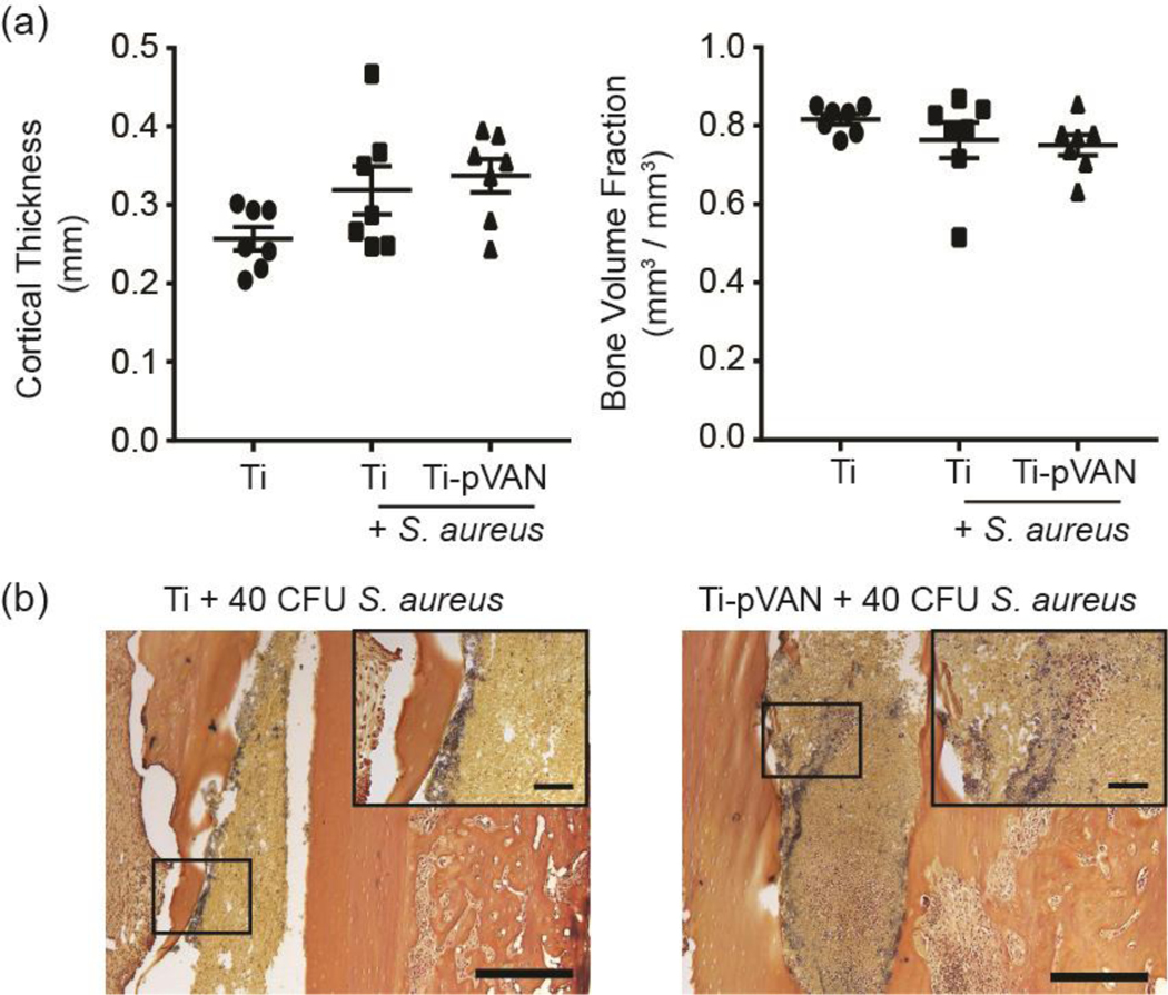
Figure 5.
Ti-pVAN pins do not eradicate S. aureus from the endosteal bone surface of infected femoral canal. (a) μ-CT quantification of cortical thickness and bone volume fraction of femur inserted with unmodified Ti6Al4V or Ti-pVAN pins, with or without 40 CFU S. aureus (n=7) at 21 days post-op. No significant difference (p>0.05, One-way ANOVA for cortical thickness & Kruskal-Wallis for bone volume fraction). (b) Gram staining of explanted femurs in the infected groups after respective pin removal at 21 days post-op (n=3). Scale bar = 500 μm; Inset: Scale bar = 100 μm.

