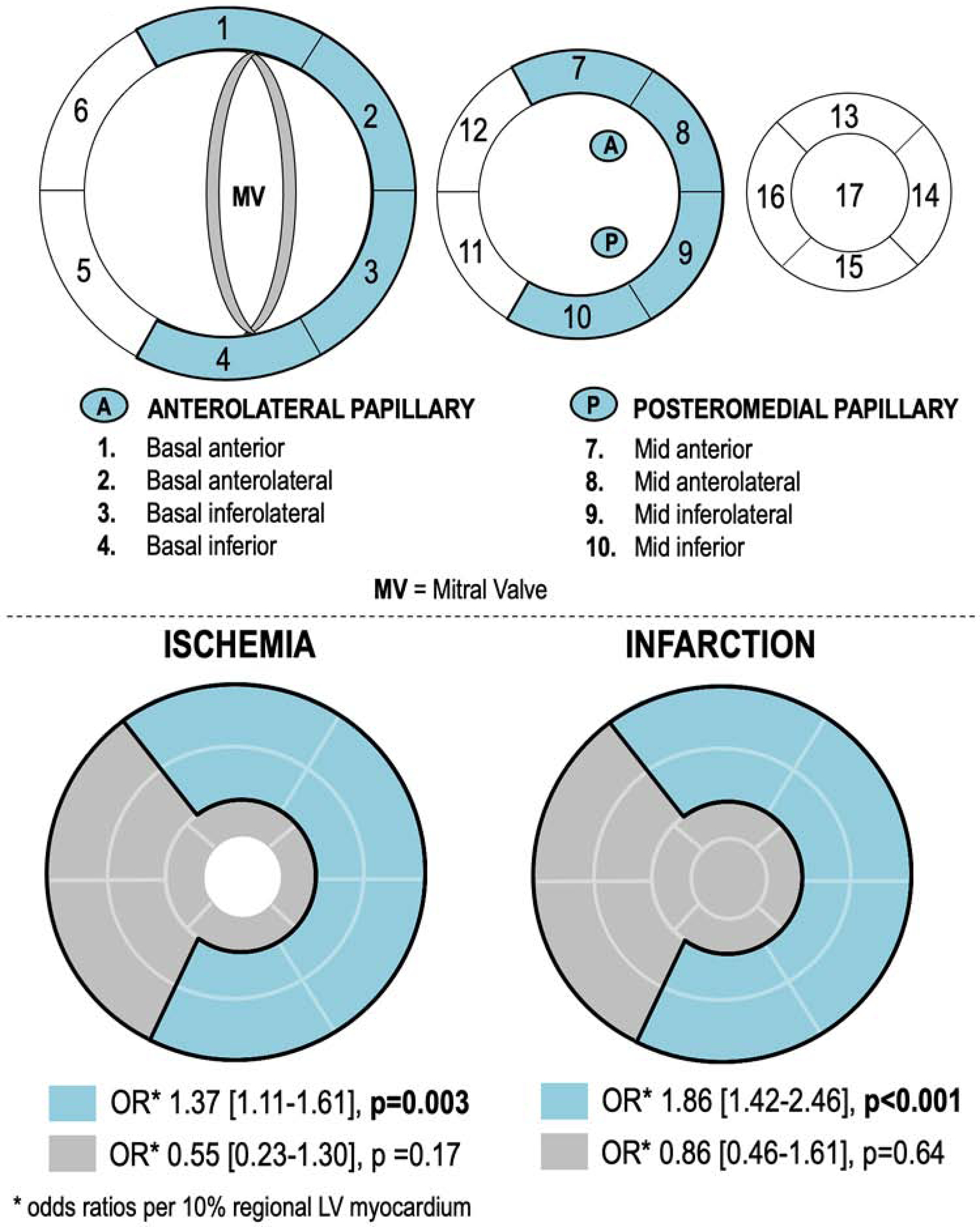
Figure 2. Mitral Apparatus Partitions.

2A. Bullseye plot (17-segment model) illustrating LV wall myocardium subtended within the mitral apparatus, which was defined as segments adjacent to the anterolateral and posteromedial papillary muscles (blue denotes sub-papillary regions [basal-mid anterior/anterolateral, inferior/inferolateral walls]).
2B. Converged bullseye plots depicting associations of FMR with LV wall ischemia (left) and infarction (right) localized to sub-papillary regions (blue) or regions anatomically distant from the papillary muscles (grey): Patients with ischemia or infarction isolated to sub-papillary regions were more likely to have advanced FMR (both p<0.01), whereas those with ischemia or infarction isolated to other regions were not (p=NS).
