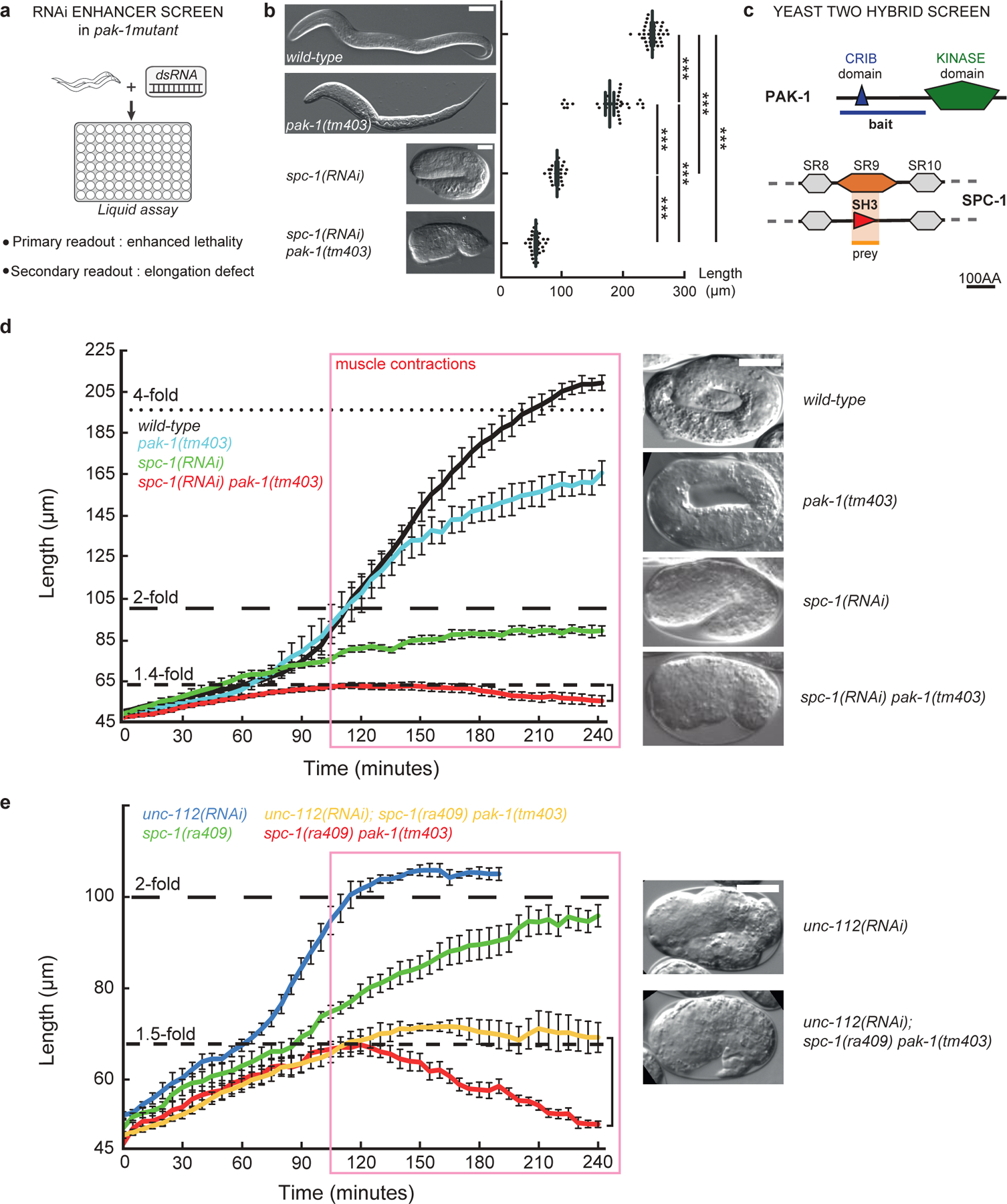
Figure 2: Loss of PAK-1 and SPC-1 triggers a muscle-dependent retraction of embryos.

(a) RNAi screen in a pak-1 mutant identified spc-1 as an enhancer (Table S1). (b) DIC micrographs of newly hatched wild-type, pak-1(tm403) (scale bar: 10 μm), spc-1(RNAi) and spc-1(RNAi) pak-1(tm403) (scale bar: 25 μm). Quantification of L1 hatchling body length: wild-type (n=38); pak-1(tm403) (n=32); spc-1(RNAi)(n=26); spc-1(RNAi) pak-1(tm40) (n=36). (c) A yeast two-hybrid screen using the PAK-1 N-term domain as a bait identified the SPC-1 SH3 domain as a prey (orange background) (Table S2). (d) Elongation profiles and corresponding terminal phenotypes of wild-type (n=5), pak-1(tm403) (n=5), spc-1(RNAi) (n=8), spc(RNAi) pak-1(tm403) (n=8). (e) Elongation profiles in a muscle defective background. unc-112(RNAi) (n=5); spc-1(RNAi) (n=8); unc-112(RNAi); pak-1(tm403) spc-1(ra409) (n=5); spc-1(RNAi) pak-1(tm403) (n=8). Right bracket (d, e), extent of retraction for spc-1(RNAi) pak-1(tm403) embryos. Scale bars, 10 μm. Error bars, SEM.
