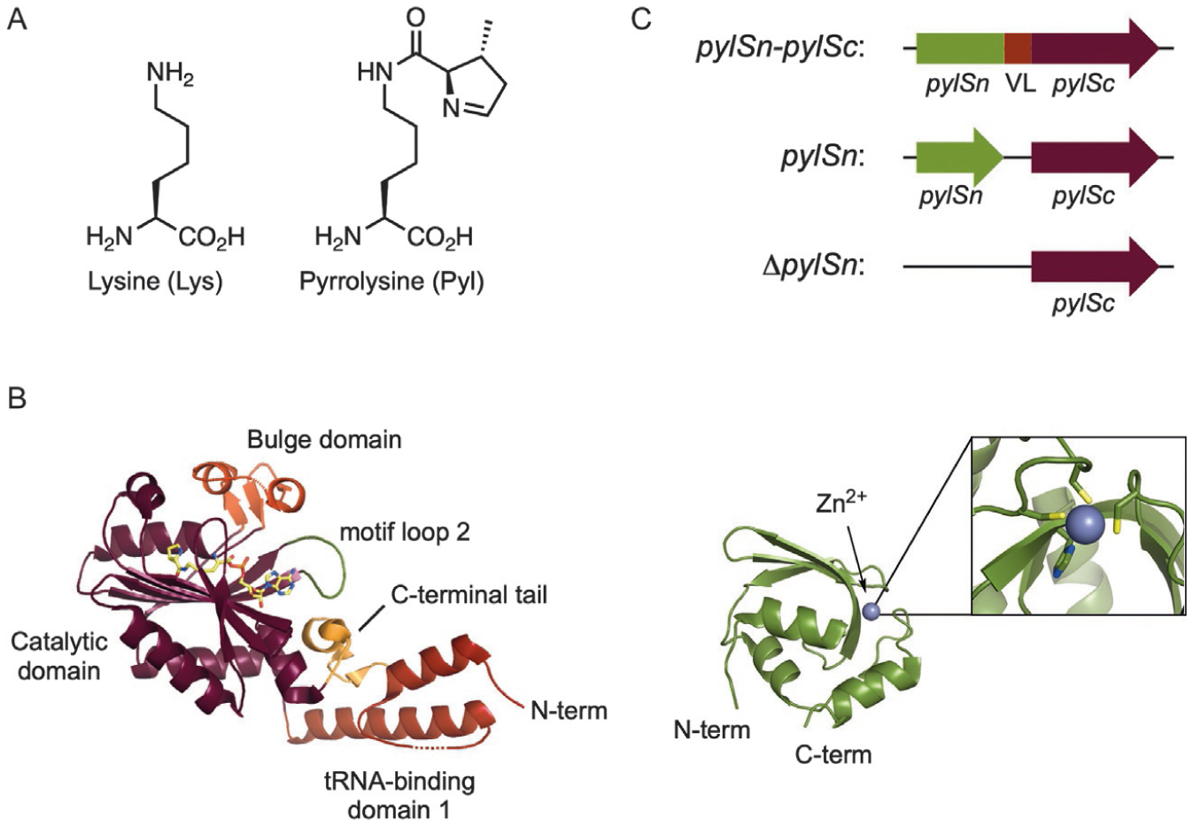
Fig. 3.

(A) The structures of l-lysine and l-pyrrolysine. (B) The structures of the C-terminal (left) and N-terminal (right) domains of PylRS from M. mazei. (PDB: 2Q7H, 5UD5) [15,35]. (C) Organization of the N-terminal (pylSn) and C-terminal (pylSc) domains of PylRS from the three defined classes (VL=variable linker).
