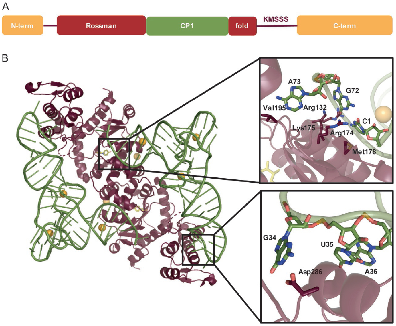
Fig. 9.

(A) Domain structure of MjTyrRS. (B) Interaction of MjTyrRS dimer with two MjtRNATyr molecules (PDBID: 1J1U) [103]. Closer look at the acceptor stem (upper box) and anticodon loop (lower box) displays the specific residues of MjTyrRS (maroon) and MjtRNATyr (green) involved in the interaction.
