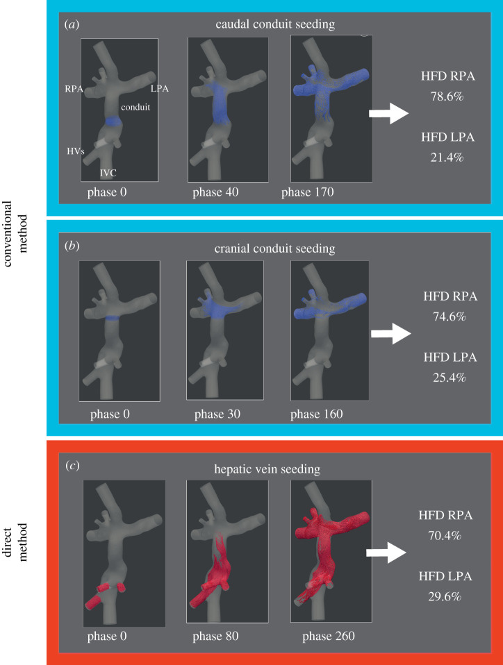
Figure 2.
The three HFD quantification approaches are shown for a typical Fontan patient. Particles were uniformly seeded from the caudal or cranial conduit (conventional method) or directly from the hepatic veins (direct method). The starting position and the trajectory of these particles over time are shown for two cardiac phases. The percentage of particles arriving at each pulmonary artery were recorded representing the HFD. IVC, inferior vena cava; HVs, hepatic veins; LPA/RPA, left/right pulmonary artery; HFD, hepatic flow distribution.

