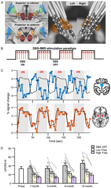
FIGURE 1:
Approximate electrode placement, functional magnetic resonance imaging (fMRI) stimulation paradigm, and clinical outcomes achieved by subthalamic nucleus (STN)-deep brain stimulation (DBS). (A) Approximate locations of electrodes are shown for all 14 Parkinson disease patients. Contacts for electrode lead placements were identified from postsurgical computed tomographic images and then projected onto the Montreal Neurological Institute brain template. The bilateral STN (orange), globus pallidus externus (blue), globus pallidus internus (GPi; green), and red nucleus (dark red) are shown. (B) We used a block-design fMRI stimulation paradigm. Stimulations to the bilateral STN were delivered during 36-second ON blocks followed by 24-second OFF blocks wherein no stimulation was delivered. Each fMRI run consisted of a total of 6 ON blocks and 6 OFF blocks. (C) Exemplar blood oxygen level–dependent (BOLD) signal responses from a single patient induced by high-frequency stimulation (130Hz) at the 12-month postsurgical treatment visit. Black lines depict expected percentage BOLD signal change. BOLD signal changes in the primary motor cortex (top panel; blue) and bilateral GPi (bottom panel; red) are also shown. (D) Bar graph illustrating the clinical outcome measure using total mean Unified Parkinson Disease Rating Scale, section III (UPDRS-III) scores in 14 patients, prior to (white bar) and 1, 3, 6, and 12 months following DBS surgery. Motor symptom scores using the UPDRS-III were measured during DBS OFF (baseline, gray bars) and DBS ON states delivered at low (yellow bars) and high (pink bars) frequencies at 1, 3, 6, and 12 months postoperatively.

