
Fig. 6. NLRX1 associates with FASTKD5 and modulates the OXPHOS pathway and HIV-1 replication in T cells.
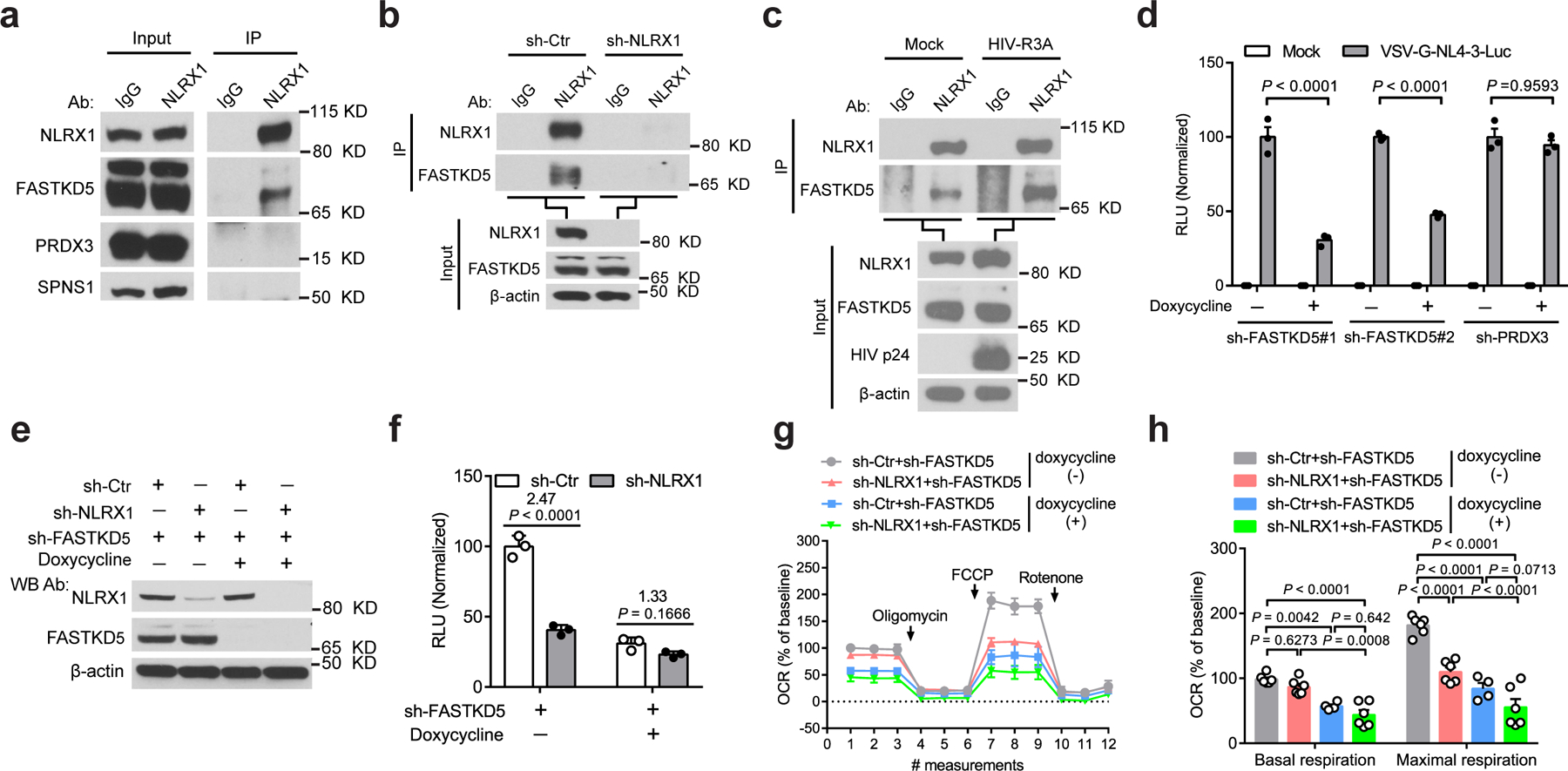
a, Interactions between NLRX1 and the mitochondrial proteins at the endogenous level. cell lysate: input; immunoprecipitated sample: IP. IgG: IP negative control.
b, NLRX1 interacted with FASTKD5 at the endogenous level. IgG: IP negative control. β-actin: loading control.
c, HIV-1 infection enhanced the association of NLRX1 and FASTKD5. HIV-1 R3A-infected Jurkat cells were subjected to IP by NLRX1 antibody or IgG at 72 hpi. β-actin: loading control.
d, RLU in FASTKD5- or PRDX3-silenced Jurkat cells at 24 hpi. n = 3 cell cultures per experiment.
e, Jurkat-sh-Ctr and Jurkat-sh-NLRX1 transduced with a lentivirus that contains a Tet-on promoter-controlled shRNA for FASTKD5 were treated with doxycycline for 72 hr or left untreated. Immunoblot shows the silencing of FASTKD5 and NLRX1. β-actin: loading control.
f, Cells were treated as described in e, followed by infection with VSV-G-NL4–3-Luc. RLU was measured at 24 hpi. n = 3 cell cultures per experiment.
g, Silencing of FASTKD5 in Jurkat-sh-Ctr and Jurkat-sh-NLRX1 was induced by doxycycline for 72 hr, followed by the infection of VSV-G-NL4–3-Luc. OCR was determined at 24 hpi. n = 6 cell cultures per experiment.
h, Basal respiration and maximal respiration. Each dot represents one cell culture.
Data are representative of three independent experiments, and panels d, f, g, and h are shown as the mean ± s.e.m. Statistical significance was tested by two-way ANOVA followed by Tukey’s (d, h) or Sidak’s (f) multiple comparisons test.