Original citation: J Clin Invest. 2020;130(6):3124–3136. https://doi.org/10.1172/JCI135026
Citation for this corrigendum: J Clin Invest. 2021;131(9):e150328. https://doi.org/10.1172/JCI150328
We identified errors in seven figure panels in this manuscript: Figures 1F, 2C, 2E, 3C, and 7E and Supplemental Figures 1E and 7A. These errors were identified as the result of a thorough review of each figure panel after discrepancies were noted by a reader after publication. When the concern was brought to our attention, we reviewed all the data for every panel in the publication. In the data review, we reexamined the original raw data for each figure. We informed the JCI of the errors in these panels and provided the raw data to the editors for their review. This information was also provided for an independent institutional evaluation. The correct data are consistent with the conclusions made in the original article. In this Corrigendum, the seven incorrect figure panels are replaced with the correct panels made from the original raw data. In addition, anti-caspase 1 antibody (catalog 22915-1-AP, Proteintech), which was omitted from the reagent list in Supplemental Table 2, has been added to the updated supplemental file.
We regret the errors.
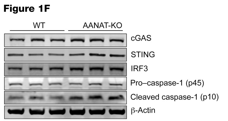
In Figure 1F the band labeled “cleaved caspase-1 (p45)” should have been labeled “cleaved caspase-1 (p10).” The correctly labeled image is below.
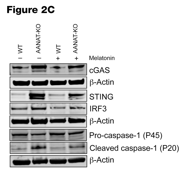
In Figure 2C, the β-actin Western blot image was inadvertently duplicated. The correct corresponding β-actin images are shown below with their respective Western blots.
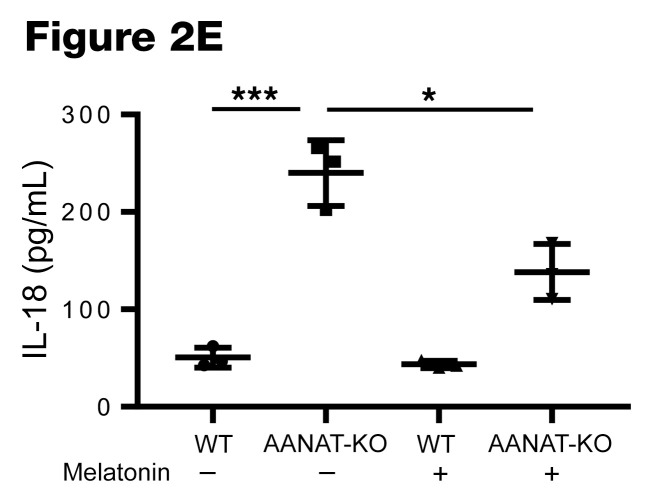
In Figure 2E, the IL-18 data were inadvertently replaced with a duplicate of the IL-1β data. The correct panel for IL-18 is shown below.
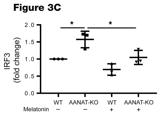
In Figure 3C, the IRF3 data were inadvertently replaced with a duplicate of the cGAS data. The correct IRF3 panel is shown below.
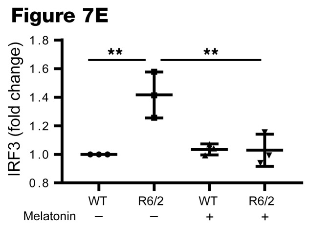
In Figure 7E, the IRF3 panel was inadvertently replaced with a duplicate of the STING panel. The correct panel for IRF3 densitometric quantification is shown below.
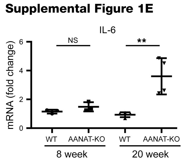
In Supplemental Figure 1E, 20-week IL-6 data were inadvertently replaced with a duplicate of the IL-1β data. The 8-week data were correct. The correct panel for IL-6 is shown below.
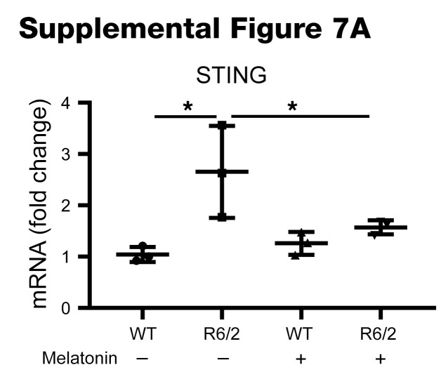
In Supplemental Figure 7A, the panel on the far right was inadvertently mislabeled as IRF3 rather than STING. The correct panel is shown below.
Version 1. 05/03/2021
Electronic publication


