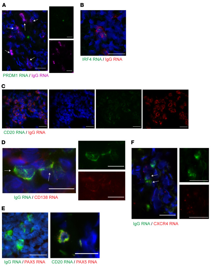
Figure 2. Cells expressing a high level of IgG RNA infiltrate in genital skin during the tissue-based immune response to HSV-2 reactivation.
(A) A healing lesion shows PRDM1 (Blimp-1) expression in many IgG RNA+ cells (40× immersion lens; scale bars: 25 μm). Result shown for 1 of 5 participants. Brightness increased for visualization. (B) IgG-producing cells, by FISH. IgG mRNA (red) and IRF4 (green) in a biopsy specimen taken during healing after HSV-2 reactivation. Note 2 cells with lower amplitude of IgG that are IRF4–. Image is 60× by confocal microscopy. Scale bar: 25 μm; result shown for 1 of 7 participants. (C) IgG RNA (red) and CD20 RNA (green) in adjacent cells without coexpression. Image is 40× by oil immersion microscopy; scale bars: 25 μm. Result shown for 1 of 5 participants. (D) IgG (green) and CD138 (red) coexpression by deconvolution microscopy (60×, single Z-stack image shown). Eight of 20 IgG RNA+ cells in this image also express CD138. Insets are single-color images of center cells. Result shown for 1 of 4 participants. (E) PAX5 RNA (red) and highly expressed IgG RNA (green) were not observed to colocalize, whereas PAX5 RNA and CD20 RNA were. Image is 40× by oil immersion microscopy. Scale bars: 25 μm; result shown for 1 of 5 participants. (F) In an active-lesion specimen, 2 cells show coexpression of CXCR4 and IgG (40× immersion lens; scale bars: 25 μm). Result shown for 1 of 3 persons. Arrows indicate cells with coexpression.

