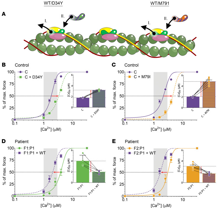
Figure 6. The experimental design and results of the reconstitution of myofibers with recombinant fsTnC.
(A) The schematic depicts the thin filament with troponin complex in which (I) endogenous fsTnC is removed from fast-twitch myofibers, followed by (II) reconstitution with exogenous fsTnC. (B and C) Normalized force-[Ca2+] relationships of myofibers from control subjects before and after reconstitution with recombinant D34Y-fsTnC (B) and M79I-fsTnC (C). Insets show the [Ca2+] at which 50% of maximal force is reached. (D and E) Normalized force-[Ca2+] relationships of myofibers from F1:P1 (D) and F2:P1 (E) before and after reconstitution with recombinant WT-fsTnC. Insets show the [Ca2+] at which 50% of maximal force is reached. The physiological [Ca2+] range is indicated by the vertical gray bar. Data are depicted as mean ± SEM.

