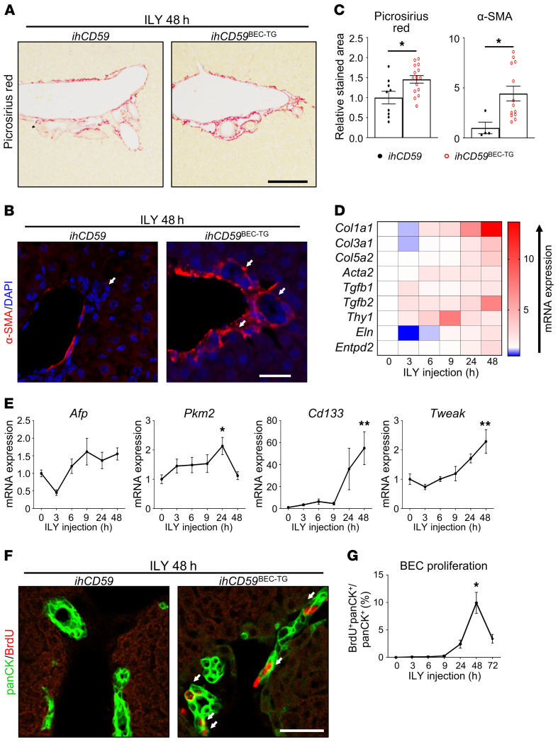
Figure 2. Acute BEC-specific injury alone triggers portal fibrogenesis and BEC proliferation.
ihCD59 and ihCD59BEC-TG mice were injected intravenously with ILY and euthanized at the indicated time points. Forty-eight hours after ILY injection, (A) Picrosirius red (scale bar: 100 μm) and (B) α-SMA (red) staining was performed. White arrows in the left panel indicate bile ducts, white arrows in the right panel indicate bile ducts surrounded by α-SMA. Scale bar: 20 μm. (C) Picrosirius red– and α-SMA–stained areas were quantified (n = 4–15 per group). (D) Expression of fibrogenesis-related genes was assessed in liver homogenates at the indicated time points after ILY administration (n = 3–7 per group). Statistical analysis is shown in Supplemental Figure 2. (E) Hepatic expression of liver regeneration–associated genes was assessed by qRT-PCR (n = 3–7 per group). (F) Mice were injected with BrdU 2 hours prior to euthanization, and panCK (green) and BrdU (red) staining was performed. White arrows indicate proliferating BECs that incorporated BrdU. (G) BrdU+panCK+ cells were quantified (n = 9–18 per group). Scale bar: 20 μm. Data represent the mean ± SEM. *P < 0.05 and **P < 0.01, compared with control ihCD59 mice, by 1-way ANOVA (E and G) and an unpaired Student’s t test (C).

