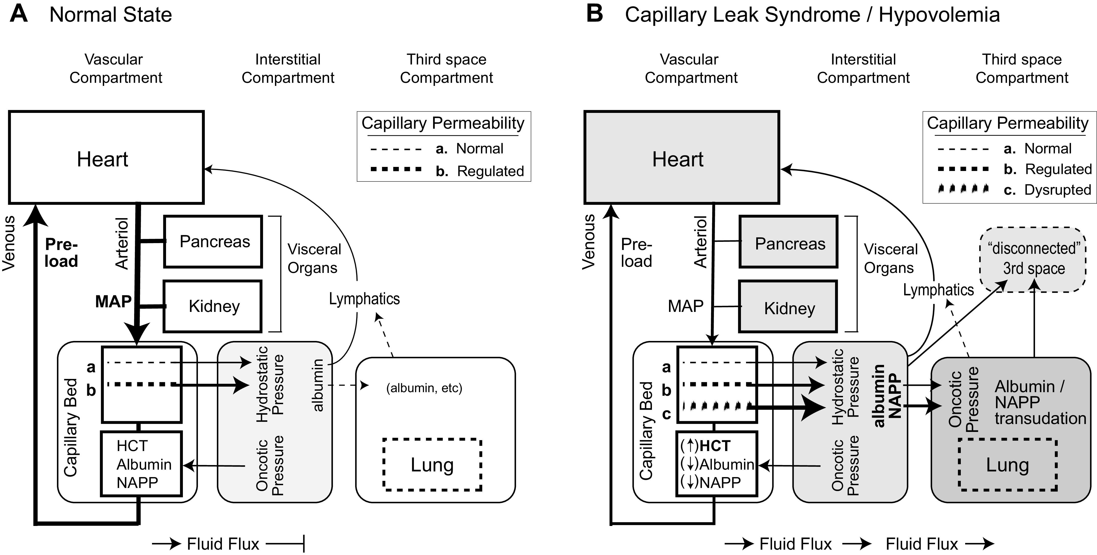
Fig. 1.

The pathophysiology of fluid sequestration: normal physiology (A) and vascular leak syndrome and hypovolemia (B). Three fluid compartments are organized as an influence diagram with the lymphatic fluid eventually returning to the heart. The key variable in B is addition of increased, large-molecule capillary permeability from disrupted endothelial barrier (c). The shaded nodes reflect sequential dysfunction resulting from disrupted capillary endothelial barrier with tissue edema in the interstitial compartment with direct effects on the lung (pulmonary edema), progressive hypovolemia with reduced blood return to the heart and hypotension, and reflex vasoconstriction and hypoperfusion of the pancreas and kidney. Albumin and proteins are lost into the 3rd space compartment, with either delayed lymphatic return or an irreversible loss to the system (disconnected 3rd space). HCT, hematocrit; MAP, mean arterial pressure; NAPP, nonalbumin plasma protein.
