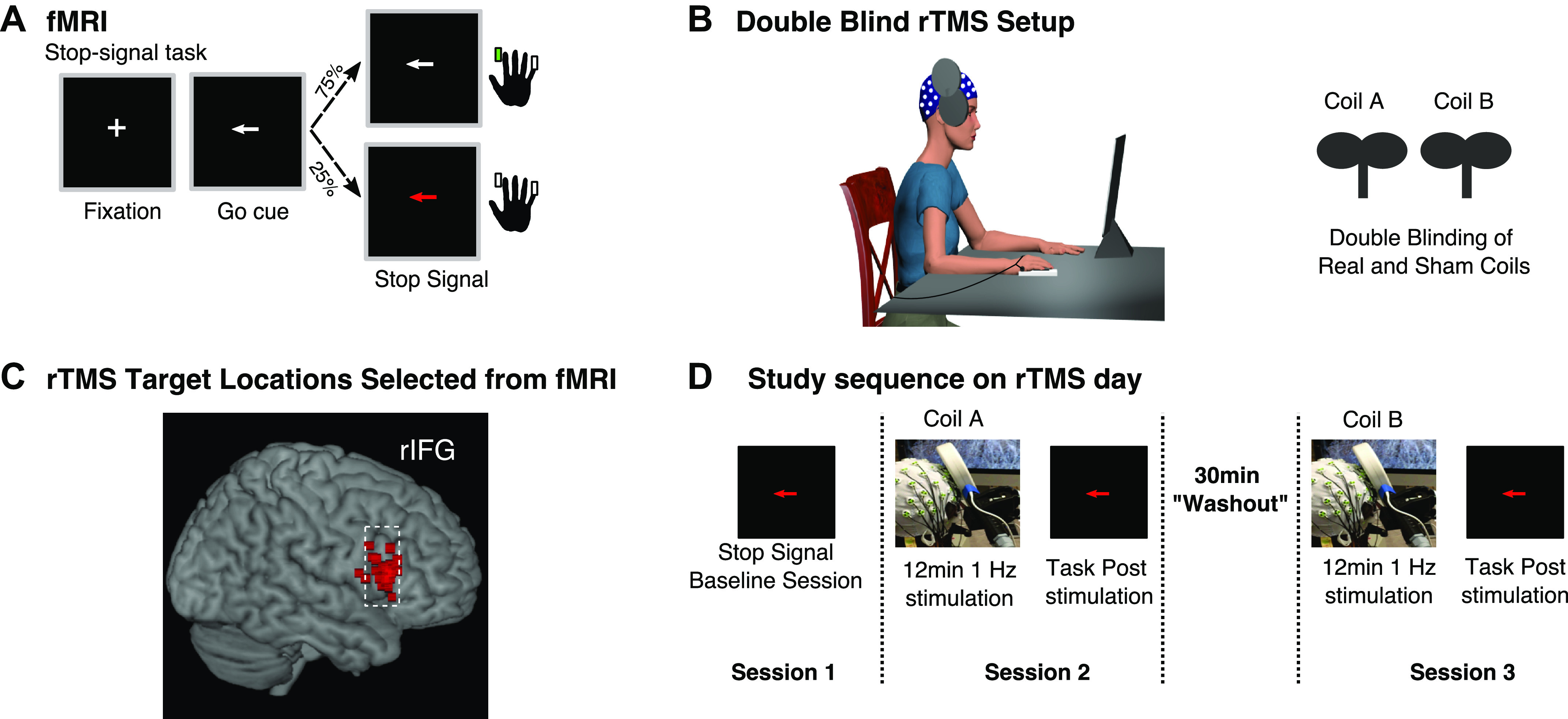
Figure 1.

Task, fMRI targeting, and EEG-transcranial magnetic stimulation (TMS) procedure. A: diagram of the stop-signal task. B: example of EEG-repetitive TMS (rTMS) setup and double-blinding procedure. C: rTMS targets selected from voxels in right inferior frontal gyrus (rIFG) with peak activation during the stop-signal task in fMRI scanner. D: sequence of events during the EEG-rTMS study visit.
