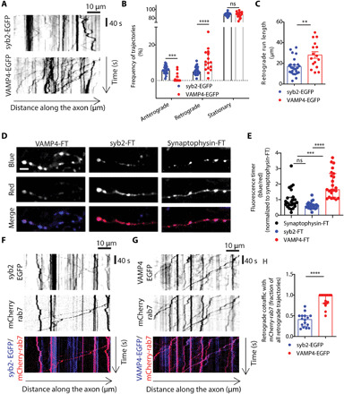
Fig. 3. VAMP4 is retrieved constitutively from axons through the endolysosomal system.

(A) Representative kymographs display the axonal mobility and directionality of traffic of VAMP4-EGFP and syb2-EGFP. (B and C) Frequency of anterograde, retrograde, or stationary trajectories (B) or the run length of retrograde trajectories (C) for both syb2-EGFP and VAMP4-EGFP. n = 25 (syb2) or n = 16 coverslips (VAMP4) from four independent preparations, ****P < 0.001 and **P = 0.0002, two-way ANOVA with Fisher’s least significant difference (B), and **P = 0.0012, unpaired t test (C). (D and E) Hippocampal neurons were transfected with syb2, synaptophysin, or VAMP4 tagged with an FT protein. (D) Representative images display FT protein fluorescence in either the blue (new protein), red (old protein), or merged channel. Scale bar, 5 μm. (E) Blue/red ratio of FT proteins. n = 24 (syb2 and synaptophysin) or n = 23 cells (VAMP4) from four independent preparations, ****P < 0.001 and ***P = 0.0004, Kruskal-Wallis test with Dunn’s multiple comparisons test. (F to H) Hippocampal neurons were transfected with mCherry-rab7 and either syb2-EGFP or VAMP4-EGFP. (F and G) Kymographs show the retrograde cotrafficking of syb2-EGFP and VAMP4-EGFP with mCherry-rab7. (H) Fraction of retrograde trajectories where syb2-EGFP or VAMP4-EGFP cotraffic with mCherry-rab7. n = 16 (syb2) or n = 18 (VAMP4) coverslips from four independent preparations, ****P < 0.001, Mann-Whitney test.
