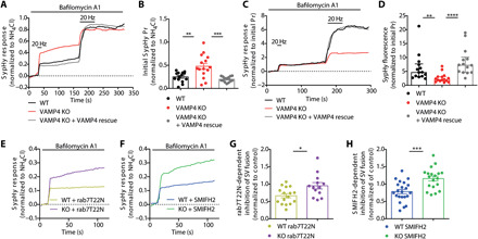
Fig. 8. VAMP4 conveys endolysosomal dysfunction into inhibition of SV fusion.

(A to D) Primary cultures of hippocampal neurons transfected with sypHy or sypHy + VAMP4-FT (VAMP4 rescue) were incubated with 1 μM bafilomycin A1 and stimulated with two 20-Hz AP trains (2 and 45 s, indicated by bar), followed by NH4Cl solution. Average traces (A) and initial evoked sypHy response normalized to the total SV pool (B). Average traces (C) and sypHy response normalized to the initial response (Pr) (D). n = 14 (wild-type, KO/rescue) or n = 15 (KO) coverslips from eight independent preparations, (B) **P = 0.0015 and ***P = 0.0001, one-way ANOVA with Tukey’s multiple comparisons test, and (D) **P = 0.0077 and ****P < 0.0001, Kruskal-Wallis test with Dunn’s multiple comparisons test. (E to H) Identical experiments to those in (A) to (D) were performed in neurons expressing either mCherry-rab7T22N or treated with 30 μM SMIFH2. (E and F) Average traces. (G and H) Inhibition of the sypHy response by either mCherry-rab7T22N (G) or SMIFH2 (H) normalized to wild-type or VAMP4 KO controls. n = 16 (wild-type/T22N), n = 13 (KO/T22N), n = 22 (wild-type/SMIFH2), and n = 19 (KO/SMIFH2) coverslips from either four (G) or three (H) independent preparations, *P = 0.012 and ***P = 0.0003, unpaired t test.
