Fig. 1.
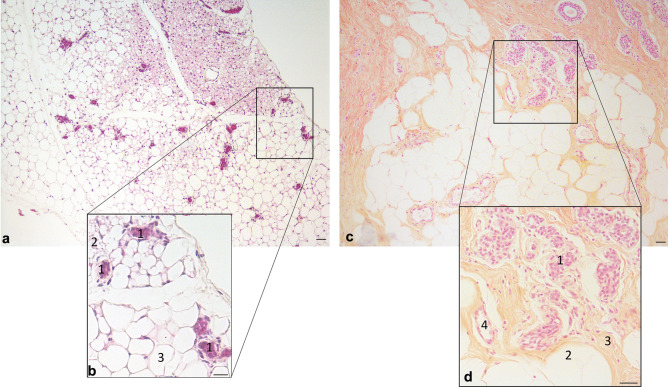
Resting Mammary Gland (light microscopy, hematoxylin and eosin staining). a: anterior mammary gland of a virgin C57BL/6 mouse containing white and brown adipocytes and ductal structures. b: magnification of squared area in A, 1: epithelial ducts; 2: brown adipocytes; 3: white adipocytes. c: Human mammary gland (resting): ductal structures and mammary stroma composed by white adipocytes and fibroblasts. d: magnification of squared area in C; 1: resting ductal structures; 2: white adipocytes; 3: fibrous stroma with fibroblasts; 4: blood vessel. Scale bars: 20μm

