Fig. 3.
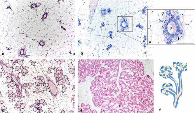
Mammary gland (mouse, 4th) during the lactation cycle. Light microscopy, hematoxylin and eosin staining. a: virgin: ductal structures infiltrating the mammary stroma mainly composed by white adipocytes. b: pregnancy (day 10): initial formation of alveolar structures arising from ductal stem cells. c: enlargement of squared area in B: 1: ductal cuboidal epithelial cells; 2: newly formed alveoli arising from ductal stem cells. 3: ductal lumen. d: Pregnancy (day 18): increased presence of alveolar structures containing large lipid vacuoles (arrow), ducts (arrowheads), accompanied by a progressive disappearance of white adipocytes. e: Lactation (day 14–15): prevalence of ductal-alveolar structures and enlarged milk-producing alveoli; adipocytes less visible. f: Cartoon representing a ductal-alveolar structure. Scale bars: 50μm

