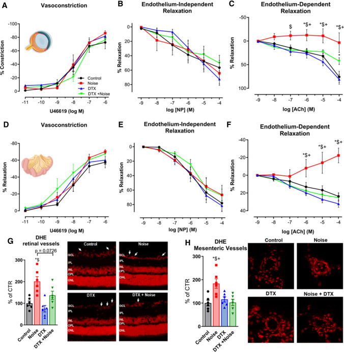
Fig. 6.
Noise-induced impairment of vasodilation and increase of oxidative stress in the retinal and mesenteric microvasculature is mostly prevented by ablation LysM+ cells. (a–c and d–f) In retinal and mesenteric arterioles, noise caused a marked degree of endothelial dysfunction (impaired ACh-response), while responses to the endothelium independent vasodilator nitroprusside (NP) and to the vasoconstrictor U46619 (thromboxane A2 agonist) remained unchanged. Ablation of LysM+ cells partially normalized the ACh-dose response relationship in retinal vessels and completely normalized it in mesenteric vessels. (g, h) DHE staining revealed an increase in ROS production in retinal and mesenteric vessels upon exposure to noise, with no effect by DTX treatment alone and partial or total prevention of the increase by DTX treatment prior to noise. Representative images of DHE-stained retinal and mesenteric cryosections are shown besides the densitometric quantification. The white arrows point to retinal vascular cross-sections. GCL ganglion cell layer; IPL inner plexiform layer; INL inner nuclear layer; OPL outer plexiform layer; ONL outer nuclear layer. Data points are measurements from individual animals (g, h) or n = 6 for (a–c) or n = 3–4 for (d–f); one-way ANOVA with Tukey’s multiple comparison test (a–c). two-way ANOVA with Bonferroni’s multiple comparison test (d–f). *P < 0.05 vs. Control; #P < 0.05 vs. Noise; $P < 0.05 vs. DTX, +P < 0.05 vs. DTX + Noise

