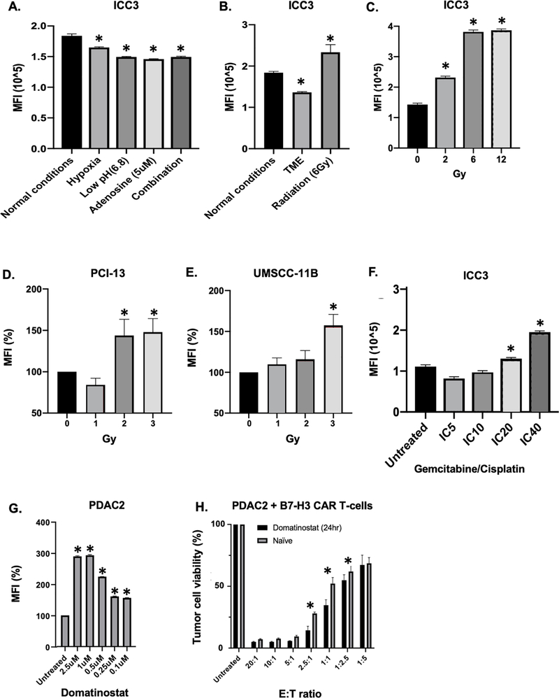
Figure 3. Modulation of B7-H3 expression on tumor cells in vitro by tumor microenvironment-like conditions, radiation and chemotherapy.
A: Intrahepatic cholangiocarcinoma ICC3 cells were cultured under different tumor microenvironment (TME)-like conditions for 6 days and subsequently B7-H3 expression was analyzed with flow cytometry using monoclonal antibody (mAb) 376.96. B: ICC3 cells were cultured either under normal conditions (normoxia, neutral pH, without adenosine) or under TME-like conditions (1% O2, pH 6.8, adenosine 5uM) for 6 days. An aliquot of cells was cultured under TME-like conditions for 3 days. These cells were then irradiated (6Gy) and kept in culture for an additional 3 days. All the cells were then analyzed for B7-H3 expression with flow cytometry using mAb 376.96. C: ICC3 cells were irradiated with different radiation doses and after 48hrs B7-H3 expression was analyzed with flow cytometry. D, E: Head and neck squamous cell carcinoma cells PCI-13 (D) and UMSCC-11B (E) were exposed to 3 fractionated doses of 2Gy, 3Gy or 4Gy radiation, with a 48hr interval between each dose. B7-H3 expression was then assessed by flow cytometry using mAb 376.96 48hrs after the last radiotherapy administration. F: Following treatment with gemcitabine and cisplatin for 72hrs, B7-H3 expression was assessed on ICC3 cells with flow cytometry using mAb 376.96. G: Following treatment with domatinostat (0.1–2.5uM) for 72hrs, B7-H3 expression was assessed on pancreatic ductal adenocarcinoma PDAC2 cells with flow cytometry using mAb 376.96. H: PDAC-2 cells were pre-treated with 1uM of domatinostat for 24hrs prior to co-culture with B7-H3 CAR T-cells. The control group (naïve) was cultured without domatinostat for 24hrs. Co-culture with B7-H3 CAR T-cells was performed for 3 days. At the end of the incubation period, viability of PDAC2 cells was assessed with an MTT assay. MFI: mean fluorescence intensity; CAR: chimeric antigen receptor; E:T: effector to target ratio. *p<0.05.

