
Figure 8.
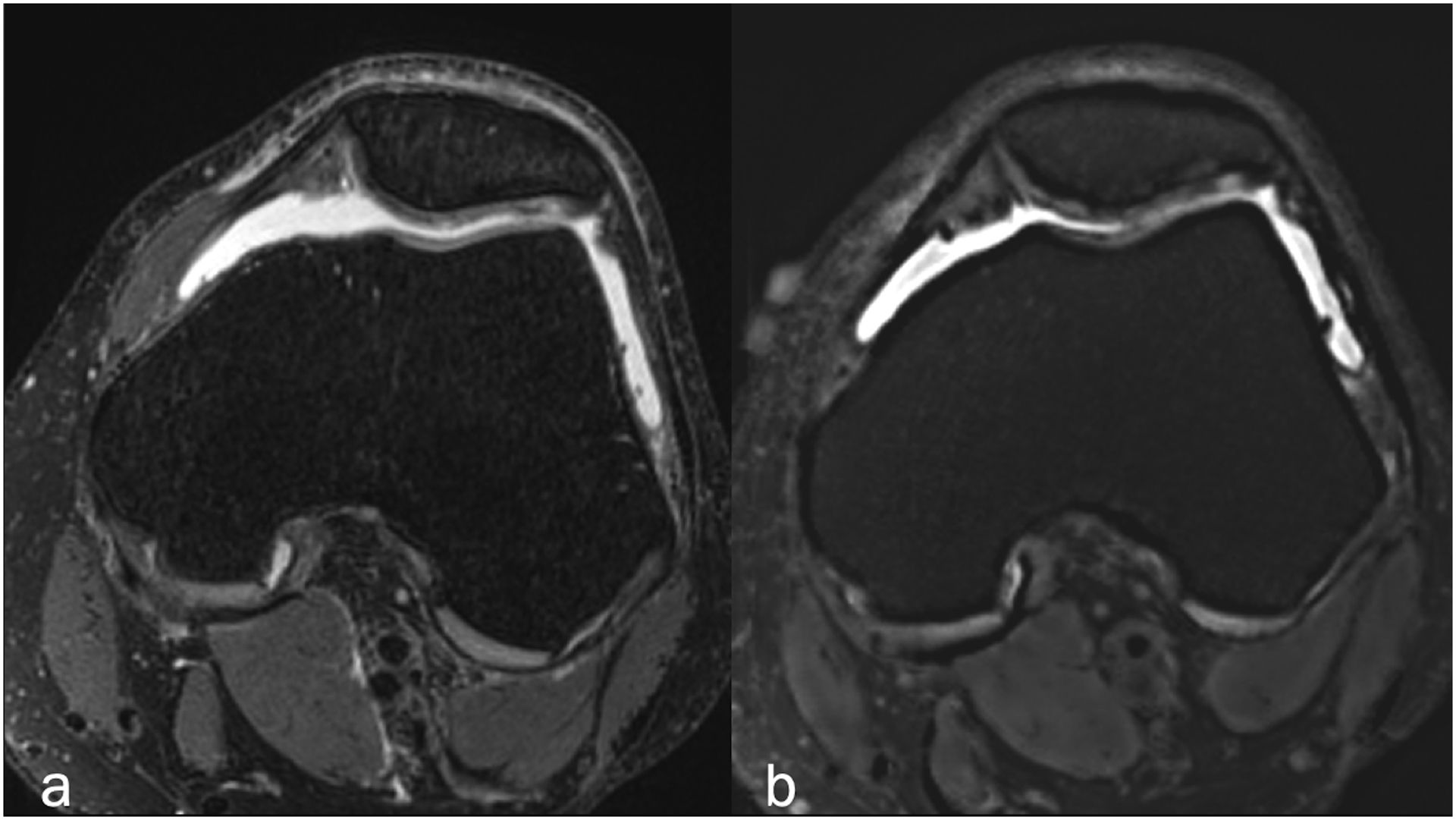
Axial (a) fat-suppressed PD-weighted, and (b) fat-suppressed synthetic MRI (AFSMRI) showing patellar cartilage with preserved thickness, normal contours and diffusely heterogeneous signal, which was comparable between the sequences. The trochlear cartilage is normal.